Copyright
©The Author(s) 2024.
World J Cardiol. Sep 26, 2024; 16(9): 531-541
Published online Sep 26, 2024. doi: 10.4330/wjc.v16.i9.531
Published online Sep 26, 2024. doi: 10.4330/wjc.v16.i9.531
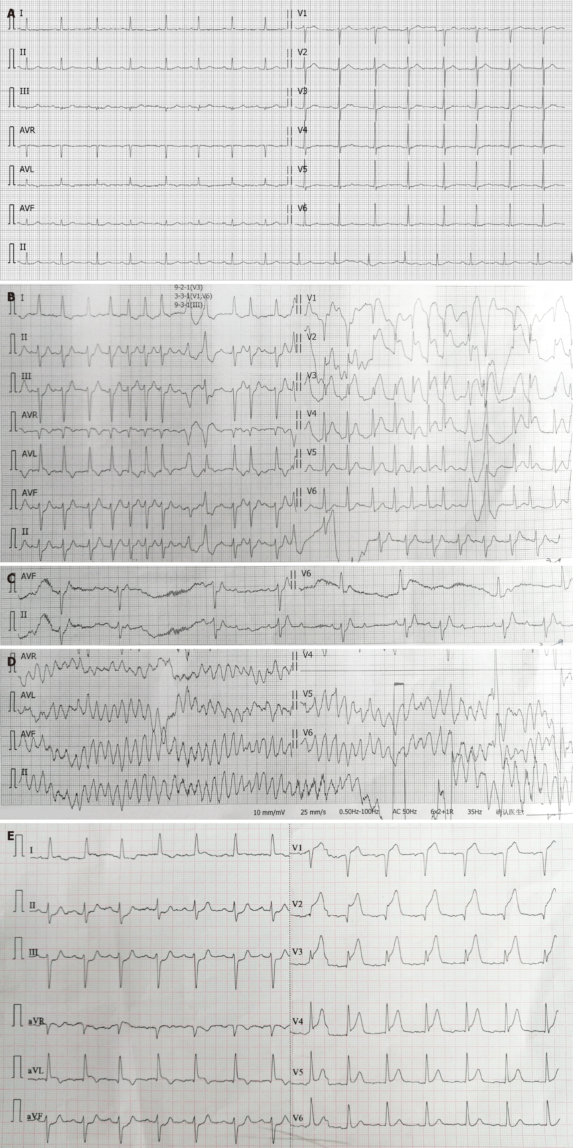
Figure 2 Transient evolution of electrocardiogram in hyperacute myocardial infarction.
A: Baseline electrocardiogram (ECG) on January 29, 2018; B and C: Initial emergency ECG at 11:38 on June 24, 2019, showing atrial fibrillation with ST-segment elevation or depression in some leads, ventricular premature beats, and spontaneous conversion of atrial fibrillation to sinus rhythm with second-degree type I atrioventricular block within one minute; D and E: ECG during syncope at 11:40 on June 24, 2019, showing torsades de pointes ventricular tachycardia (TdP), with TdP lasting approximately ten seconds before spontaneous conversion to sinus rhythm with ST-segment elevation or depression in some leads.
- Citation: She LQ, Gao DK, Hong L, Tian Y, Wang HZ, Huang S. Intracoronary thrombolysis combined with drug balloon angioplasty in a young ST-segment elevation myocardial infarction patient: A case report. World J Cardiol 2024; 16(9): 531-541
- URL: https://www.wjgnet.com/1949-8462/full/v16/i9/531.htm
- DOI: https://dx.doi.org/10.4330/wjc.v16.i9.531


