Copyright
©The Author(s) 2024.
World J Cardiol. Sep 26, 2024; 16(9): 522-530
Published online Sep 26, 2024. doi: 10.4330/wjc.v16.i9.522
Published online Sep 26, 2024. doi: 10.4330/wjc.v16.i9.522
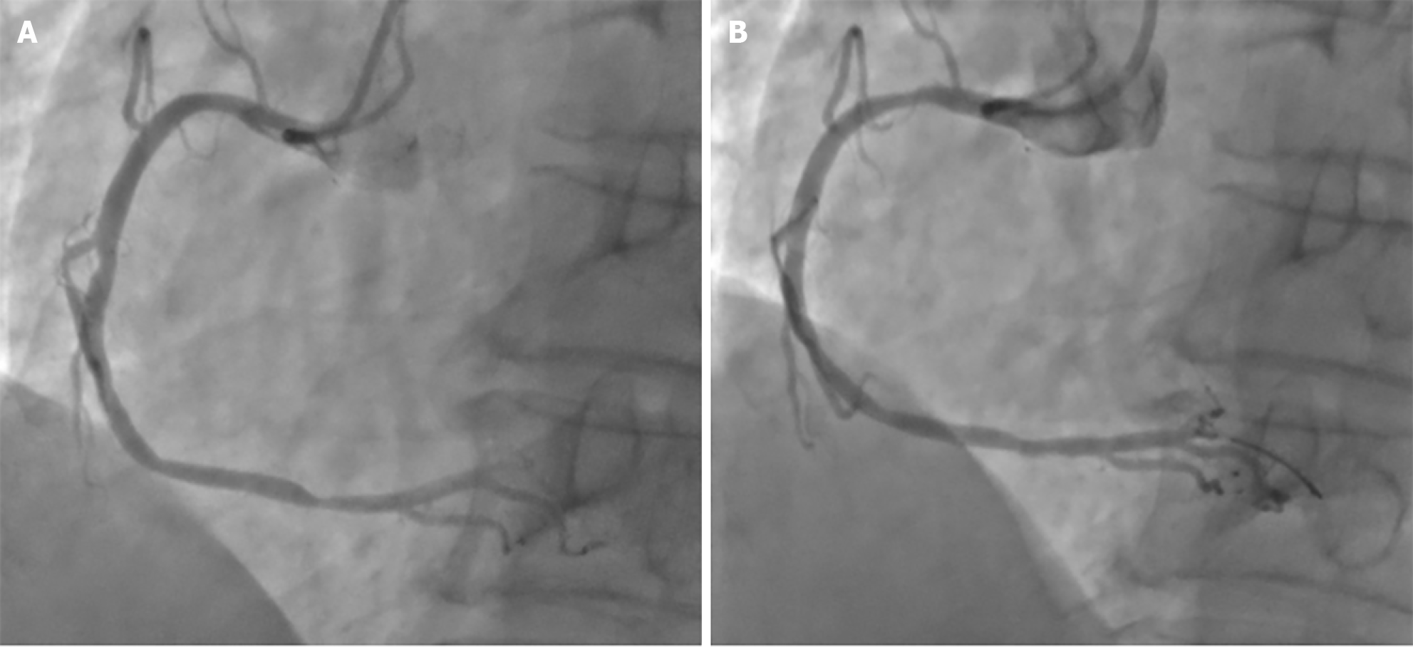
Figure 3 Coronary angiography, right coronary artery.
A: Before surgery, the stenosis in the proximal and middle segments was about 50%, the stenosis in the middle segment was limited by 50%, and the stenosis in the middle and middle segments was 40%-50%. Localized eccentric stenosis in the distal segment is about 80%. Visible posterior descending artery and posterior branch of left ventricle emissions; B: After drug balloon dilation percutaneous transluminal coronary angioplasty treatment, the lumen showed significant improvement after dilation, with visible A-type dissection and thrombolysis in myocardial infarction III blood flow.
- Citation: Zeng J, Zhao Y, Gao D, Lu X, Dong JJ, Liu YB, Shen B. Medical appraisal of Chinese military aircrew with abnormal results of coronary computed tomographic angiography. World J Cardiol 2024; 16(9): 522-530
- URL: https://www.wjgnet.com/1949-8462/full/v16/i9/522.htm
- DOI: https://dx.doi.org/10.4330/wjc.v16.i9.522


