Copyright
©The Author(s) 2024.
World J Cardiol. Sep 26, 2024; 16(9): 522-530
Published online Sep 26, 2024. doi: 10.4330/wjc.v16.i9.522
Published online Sep 26, 2024. doi: 10.4330/wjc.v16.i9.522
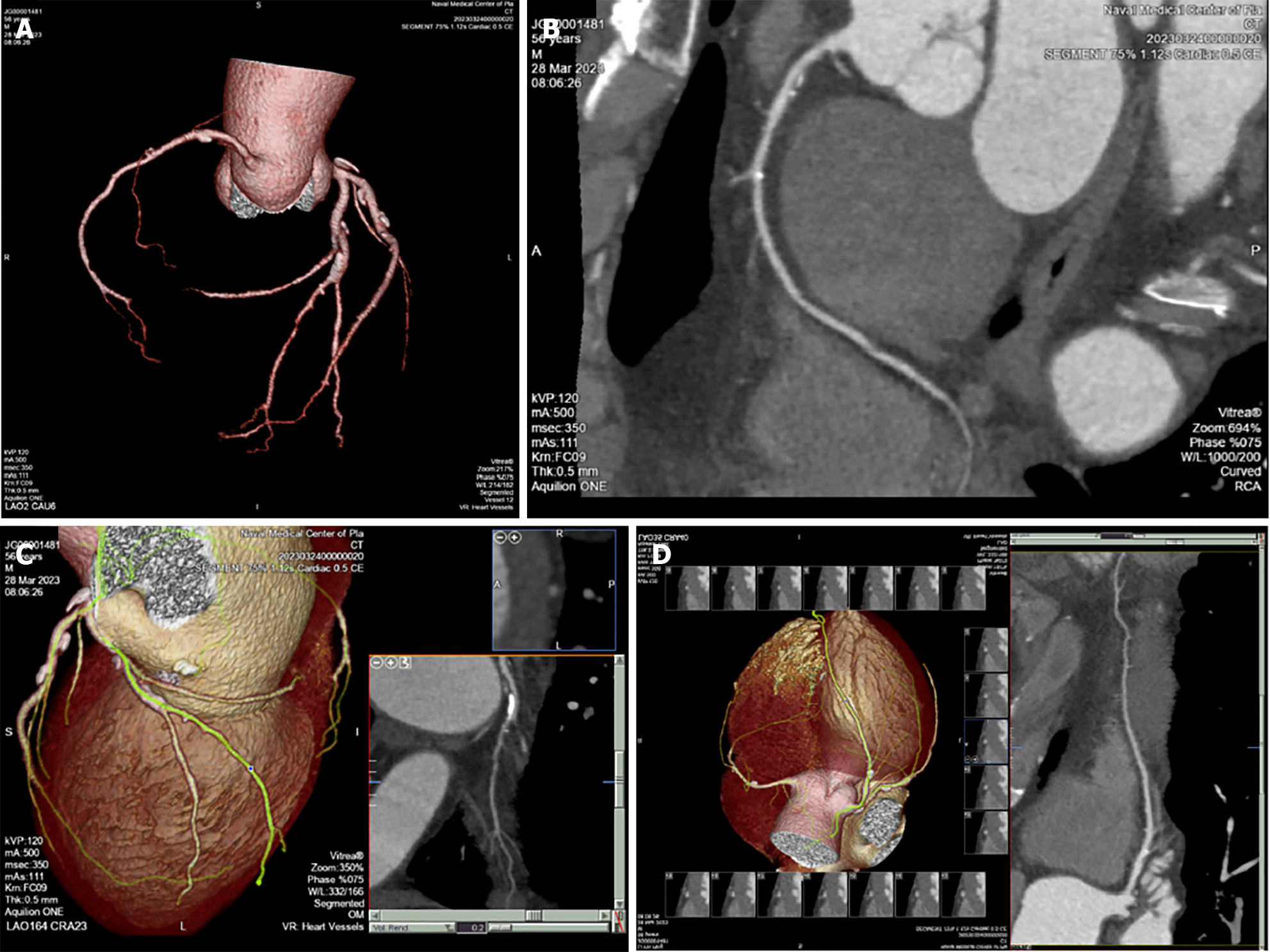
Figure 1 Transport pilot coronary computed tomographic angiography examination reveals coronary artery stenosis.
A: All coronary artery images show mixed plaques with four vessel lesions and mild to moderate stenosis of the lumen; B: Right coronary artery, mixed plaques in the proximal segment, calcified plaques in the middle segment, soft plaques in the distal segment, and mild to moderate narrowing of the lumen; C: Obtuse marginal branch mixed plaques in the proximal and middle segments, with mild to moderate narrowing of the lumen; D: Left anterior descending of coronary artery, mixed plaques in the proximal and middle segments, with mild to moderate narrowing of the lumen.
- Citation: Zeng J, Zhao Y, Gao D, Lu X, Dong JJ, Liu YB, Shen B. Medical appraisal of Chinese military aircrew with abnormal results of coronary computed tomographic angiography. World J Cardiol 2024; 16(9): 522-530
- URL: https://www.wjgnet.com/1949-8462/full/v16/i9/522.htm
- DOI: https://dx.doi.org/10.4330/wjc.v16.i9.522


