Copyright
©The Author(s) 2024.
World J Cardiol. Sep 26, 2024; 16(9): 496-501
Published online Sep 26, 2024. doi: 10.4330/wjc.v16.i9.496
Published online Sep 26, 2024. doi: 10.4330/wjc.v16.i9.496
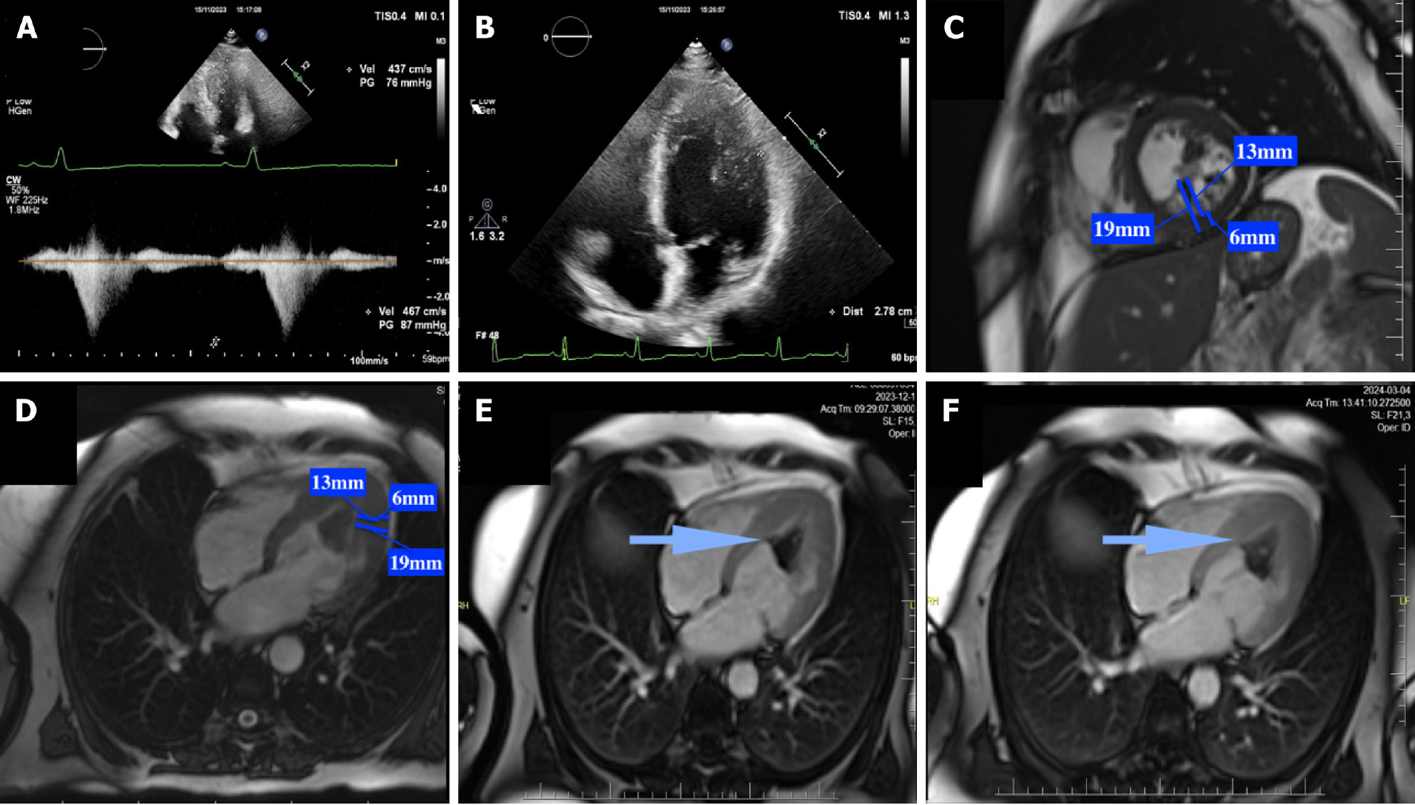
Figure 2 Images from transthoracic echocardiography and cardiac magnetic resonance imaging of a 58-year-old woman with coexistence of left ventricle hypertrophy and hypertrabeculation leading to intracavitary obstruction with heart failure symptoms, and left ventricle thrombus formation with subsequent transient ischemic attack.
A: Pulse-wave Doppler from apical 4-chamber transthoracic echocardiography (TTE) view showing intracavitary obstruction at the level of the papillary muscles (maximal resting gradient of 76 mmHg); B: Left ventricle (LV) apical hypertrophy in apical 4-chamber view in TTE; C and D: LV hypertrabeculation in cardiac magnetic resonance short axis view and 4-chamber view (ratio of thickness of non-compacted to compacted layers 2.1: 13 mm and 6 mm); E and F: LV thrombus entering between the LV trabeculae and recesses in short axis view and 4-chamber view (blue arrow).
- Citation: Przytuła N, Dziewięcka E, Winiarczyk M, Graczyk K, Stępień A, Rubiś P. Hypertrophic cardiomyopathy and left ventricular non-compaction: Distinct diseases or variant phenotypes of a single condition? World J Cardiol 2024; 16(9): 496-501
- URL: https://www.wjgnet.com/1949-8462/full/v16/i9/496.htm
- DOI: https://dx.doi.org/10.4330/wjc.v16.i9.496


