Copyright
©The Author(s) 2024.
World J Cardiol. Mar 26, 2024; 16(3): 161-167
Published online Mar 26, 2024. doi: 10.4330/wjc.v16.i3.161
Published online Mar 26, 2024. doi: 10.4330/wjc.v16.i3.161
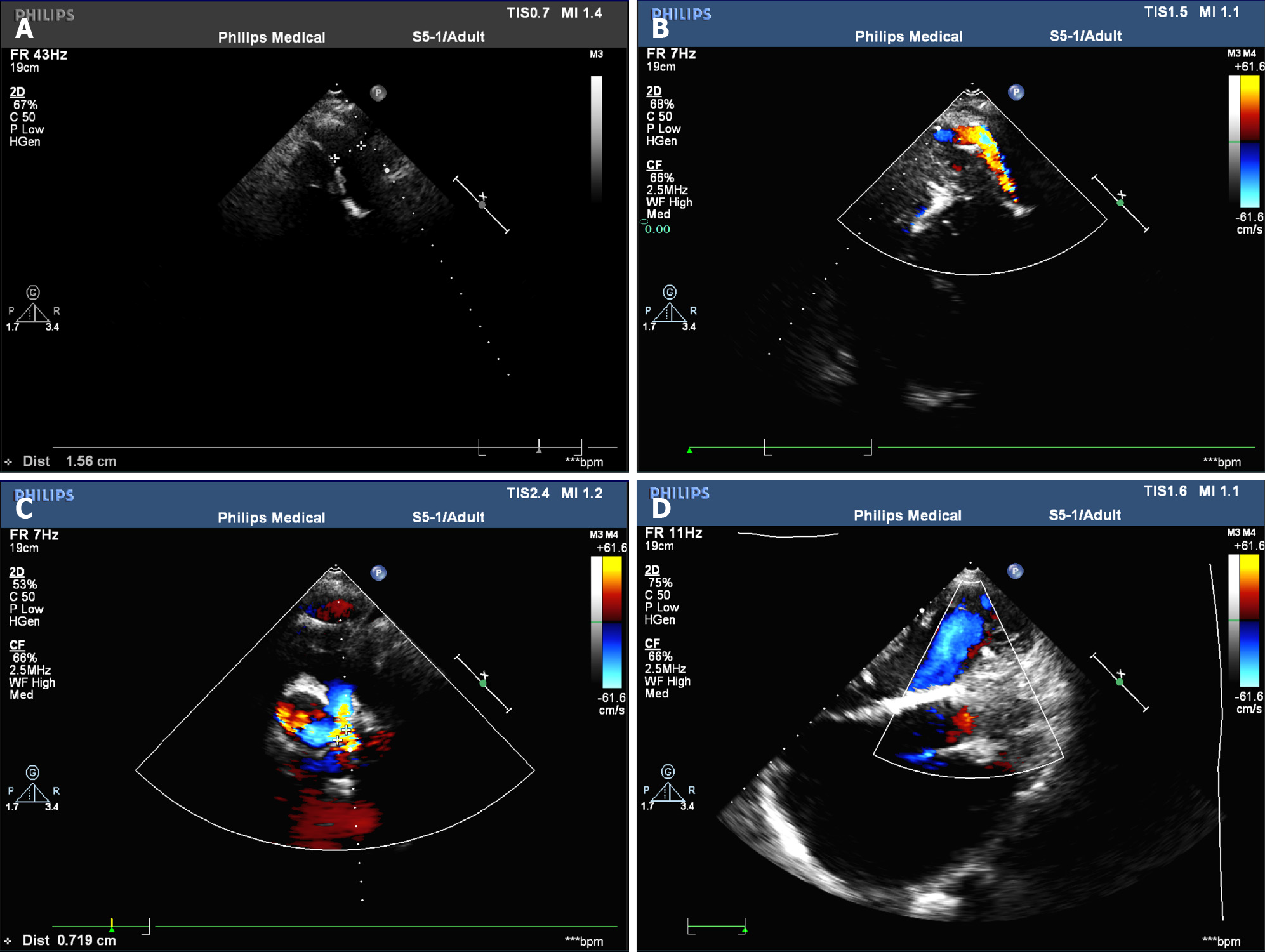
Figure 2 Preoperative echocardiography.
A and B: Artificial blood vessel shadow and blood flow signal can be seen between the descending aorta and the left pulmonary artery; C: Moderate to severe tricuspid regurgitation can be seen during cardiac systole; D: No significant residual shunt was observed at the interventricular septal level.
- Citation: Li ZH, Lou L, Chen YX, Shi W, Zhang X, Yang J. Severe hypoxemia after radiofrequency ablation for atrial fibrillation in palliatively repaired tetralogy of Fallot: A case report. World J Cardiol 2024; 16(3): 161-167
- URL: https://www.wjgnet.com/1949-8462/full/v16/i3/161.htm
- DOI: https://dx.doi.org/10.4330/wjc.v16.i3.161


