Copyright
©The Author(s) 2024.
World J Cardiol. Oct 26, 2024; 16(10): 595-603
Published online Oct 26, 2024. doi: 10.4330/wjc.v16.i10.595
Published online Oct 26, 2024. doi: 10.4330/wjc.v16.i10.595
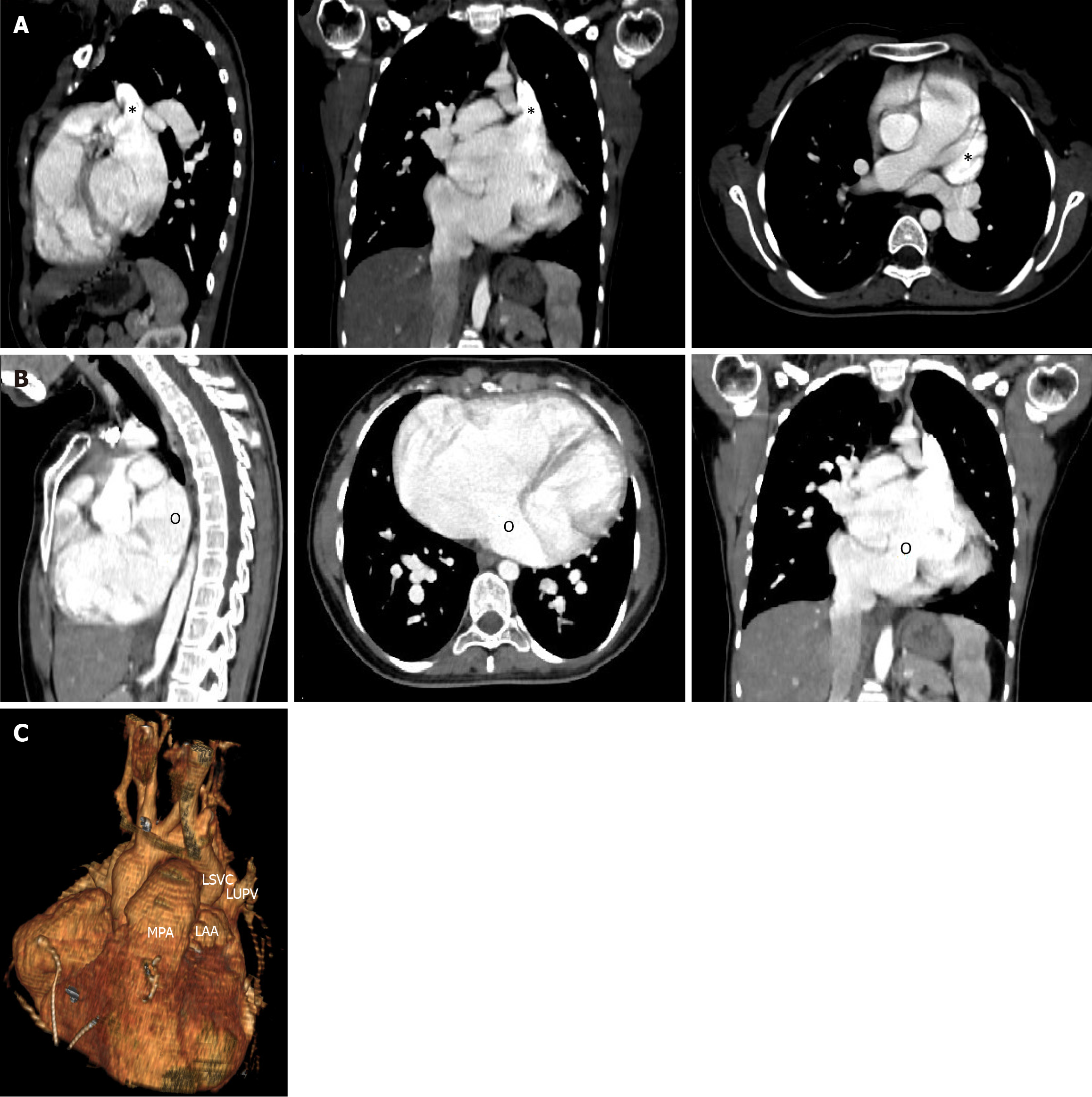
Figure 4 Preoperative computed tomography angiogram.
A: The insertion site of the left superior vena cava (LSVC) is shown by an asterisk in the axial, sagittal and coronal views; B: The ostium of the coronary sinus indicated by the letter (o) is shown in the same three views; C: A three-dimensional reconstruction of the LSCV as it enters the left atrium. LSVC: Left superior vena cava; MPA: Main pulmonary artery; LAA: Left atrial appendage; LUPV: Left upper pulmonary vein.
- Citation: Bitar F, Bulbul Z, Jassar Y, Zareef R, Abboud J, Arabi M, Bitar FF. Unroofed coronary sinus, left-sided superior vena cava and mitral insufficiency: A case report and review of the literature. World J Cardiol 2024; 16(10): 595-603
- URL: https://www.wjgnet.com/1949-8462/full/v16/i10/595.htm
- DOI: https://dx.doi.org/10.4330/wjc.v16.i10.595


