Copyright
©The Author(s) 2024.
World J Cardiol. Oct 26, 2024; 16(10): 595-603
Published online Oct 26, 2024. doi: 10.4330/wjc.v16.i10.595
Published online Oct 26, 2024. doi: 10.4330/wjc.v16.i10.595
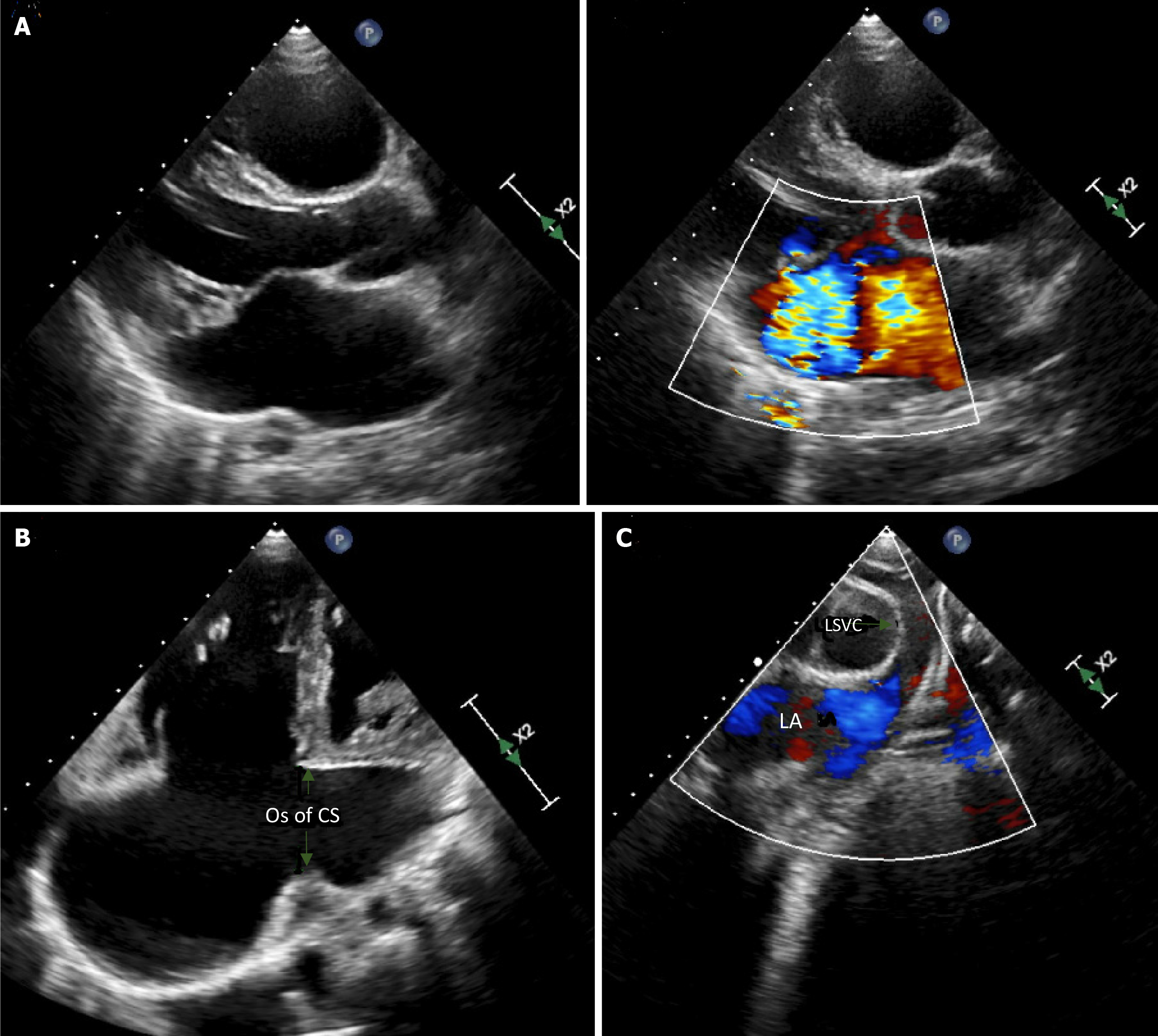
Figure 2 Echocardiographic imaging of the mitral valve.
A: A parasternal long axis view of the mitral valve showing myxomatous prolapsing leaflets and severe mitral regurgitation; B: A modified 4 chamber view, with posterior angulation showing the dilated ostium of the coronary sinus; C: Modified supra coronal cut from the supra-sternal window showing a left superior vena cava draining into the left atrium. OS: Ostium; CS: Coronary sinus; LSVC: Left superior vena cava; LA: Left atrium.
- Citation: Bitar F, Bulbul Z, Jassar Y, Zareef R, Abboud J, Arabi M, Bitar FF. Unroofed coronary sinus, left-sided superior vena cava and mitral insufficiency: A case report and review of the literature. World J Cardiol 2024; 16(10): 595-603
- URL: https://www.wjgnet.com/1949-8462/full/v16/i10/595.htm
- DOI: https://dx.doi.org/10.4330/wjc.v16.i10.595


