Copyright
©The Author(s) 2023.
World J Cardiol. Apr 26, 2023; 15(4): 119-141
Published online Apr 26, 2023. doi: 10.4330/wjc.v15.i4.119
Published online Apr 26, 2023. doi: 10.4330/wjc.v15.i4.119
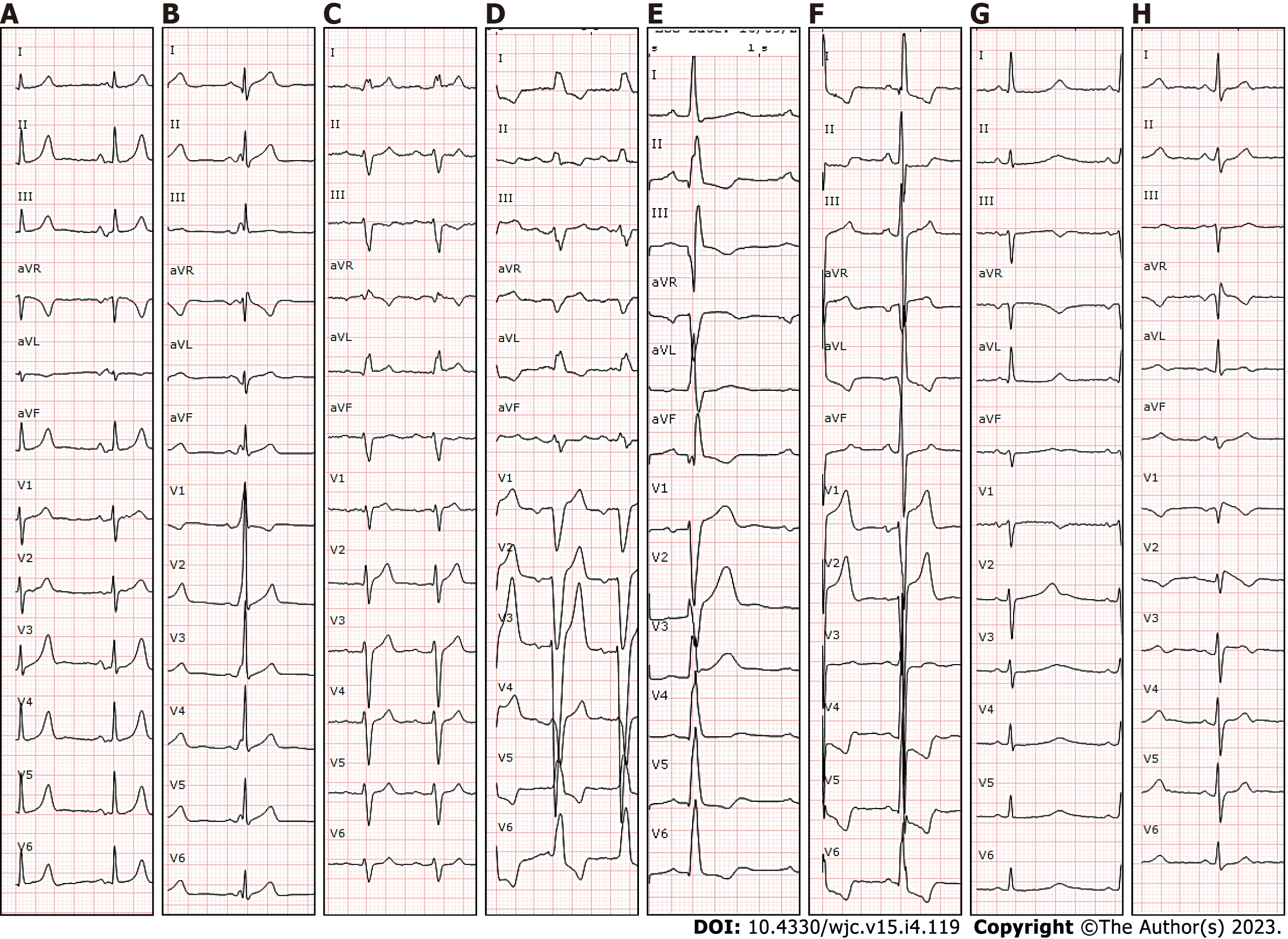
Figure 1 Examples of pathological electrocardiogram that should lead to suspicion of an arrhythmic origin of the syncope.
A: Bayes Syndrome (biphasic p wave in inferior leads compatible with interatrial block, which is related with atrial arrhythmias); B: Pre-excitation syndrome; C: Long PR interval and left anterior fascicular hemiblock; D: Left bundle branch block; E: Inferior necrosis (Q waves); F: Hypertrophic cardiomyopathy; G: Long QT syndrome; H: Brugada syndrome. Suspected supraventricular tachycardia (A and B), suspected atrioventricular block (C and D), suspected ventricular tachycardia (E and F), and suspected polymorphic ventricular tachycardia (G and H).
- Citation: Francisco Pascual J, Jordan Marchite P, Rodríguez Silva J, Rivas Gándara N. Arrhythmic syncope: From diagnosis to management. World J Cardiol 2023; 15(4): 119-141
- URL: https://www.wjgnet.com/1949-8462/full/v15/i4/119.htm
- DOI: https://dx.doi.org/10.4330/wjc.v15.i4.119


