Copyright
©The Author(s) 2023.
World J Cardiol. Dec 26, 2023; 15(12): 649-654
Published online Dec 26, 2023. doi: 10.4330/wjc.v15.i12.649
Published online Dec 26, 2023. doi: 10.4330/wjc.v15.i12.649
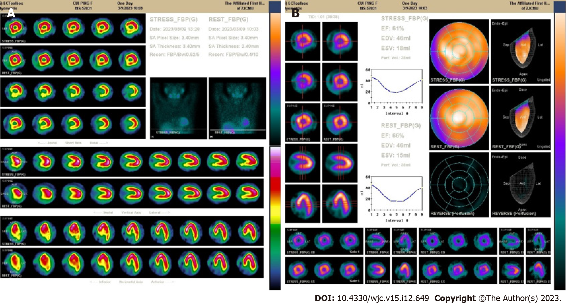
Figure 3 Results of emission computed tomography.
A: The inferior wall basal segment, inferior lateral wall basal segment, inferior septal wall middle segment, and basal segment of the left ventricle had a small-to-medium range of moderate myocardial blood flow perfusion reduction; B: Normal left ventricular systolic function without abnormal wall motion with a left ventricular ejection fraction of 66%. The load left ventricular ejection fraction was 61%.
- Citation: Zhou YP, Wang LL, Qiu YG, Huang SW. R-I subtype single right coronary artery with congenital absence of left coronary system: A case report. World J Cardiol 2023; 15(12): 649-654
- URL: https://www.wjgnet.com/1949-8462/full/v15/i12/649.htm
- DOI: https://dx.doi.org/10.4330/wjc.v15.i12.649


