Copyright
©The Author(s) 2022.
World J Cardiol. Apr 26, 2022; 14(4): 260-265
Published online Apr 26, 2022. doi: 10.4330/wjc.v14.i4.260
Published online Apr 26, 2022. doi: 10.4330/wjc.v14.i4.260
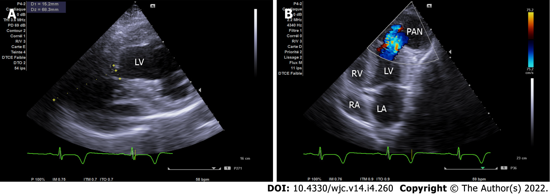
Figure 2 Transthoracic echocardiogram.
A: Modified short axis view with the pseudoaneurysm dimensions (width of the neck was 15 mm and the maximal internal diameter of the aneurysmal sac was 68 mm with a neck to sac ratio of less than 0.5); B: Apical modified 4/5 chamber view showing bidirectional shunt through the left ventricular wall.
- Citation: Jallal H, Belabes S, Khatouri A. Uncommon post-infarction pseudoaneurysms: A case report. World J Cardiol 2022; 14(4): 260-265
- URL: https://www.wjgnet.com/1949-8462/full/v14/i4/260.htm
- DOI: https://dx.doi.org/10.4330/wjc.v14.i4.260


