Copyright
©The Author(s) 2022.
World J Cardiol. Apr 26, 2022; 14(4): 206-219
Published online Apr 26, 2022. doi: 10.4330/wjc.v14.i4.206
Published online Apr 26, 2022. doi: 10.4330/wjc.v14.i4.206
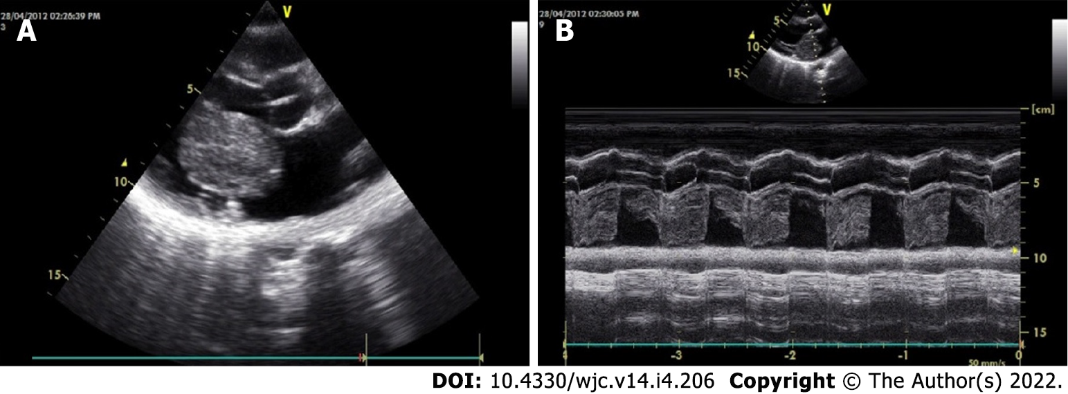
Figure 10 Echocardiographic features of myxoma.
A, B: Transthoracic echocardiography 2D (A) and M-mode (B) shows a large polypoid mass in the left atrial cavity attached to the interatrial septum by means of a stalk (not visualized here) and protruding into the left ventricular cavity across the mitral valve in diastole.
- Citation: Islam AKMM. Cardiac myxomas: A narrative review. World J Cardiol 2022; 14(4): 206-219
- URL: https://www.wjgnet.com/1949-8462/full/v14/i4/206.htm
- DOI: https://dx.doi.org/10.4330/wjc.v14.i4.206


