Copyright
©The Author(s) 2022.
World J Cardiol. Feb 26, 2022; 14(2): 64-82
Published online Feb 26, 2022. doi: 10.4330/wjc.v14.i2.64
Published online Feb 26, 2022. doi: 10.4330/wjc.v14.i2.64
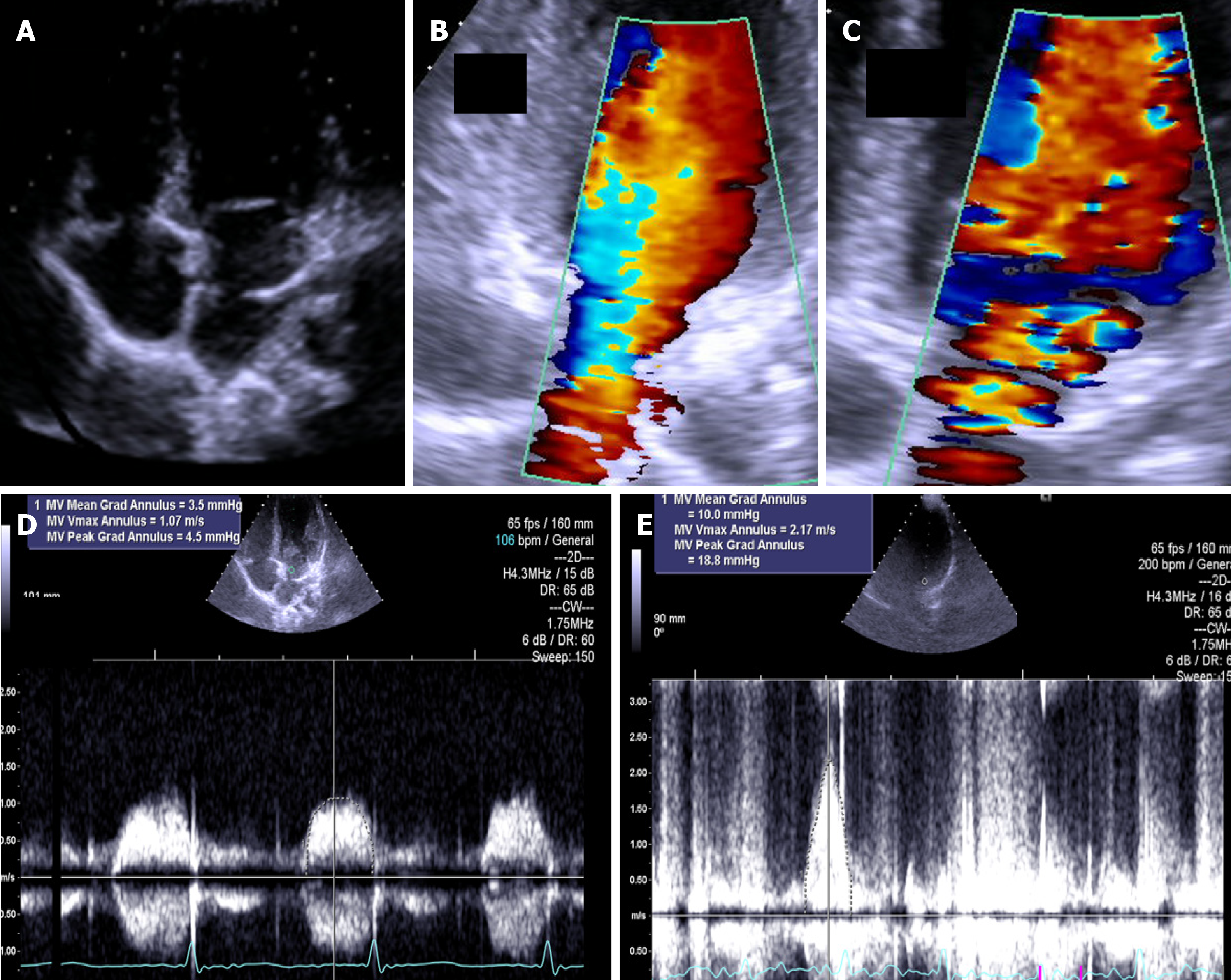
Figure 7 The exercise Doppler data in conjunction with the exercise data and clinical data led us to keep the patient in close clinical follow-up.
A: Intraauricular septum in “cor triatriatrium”; B: Color flow before exercise; C: Color flow at peak exercise; D: CW flow before exercise; E: CW flow at peak exercise.
- Citation: Cotrim CA, Café H, João I, Cotrim N, Guardado J, Cordeiro P, Cotrim H, Baquero L. Exercise stress echocardiography: Where are we now? World J Cardiol 2022; 14(2): 64-82
- URL: https://www.wjgnet.com/1949-8462/full/v14/i2/64.htm
- DOI: https://dx.doi.org/10.4330/wjc.v14.i2.64


