Copyright
©The Author(s) 2022.
World J Cardiol. Oct 26, 2022; 14(10): 546-556
Published online Oct 26, 2022. doi: 10.4330/wjc.v14.i10.546
Published online Oct 26, 2022. doi: 10.4330/wjc.v14.i10.546
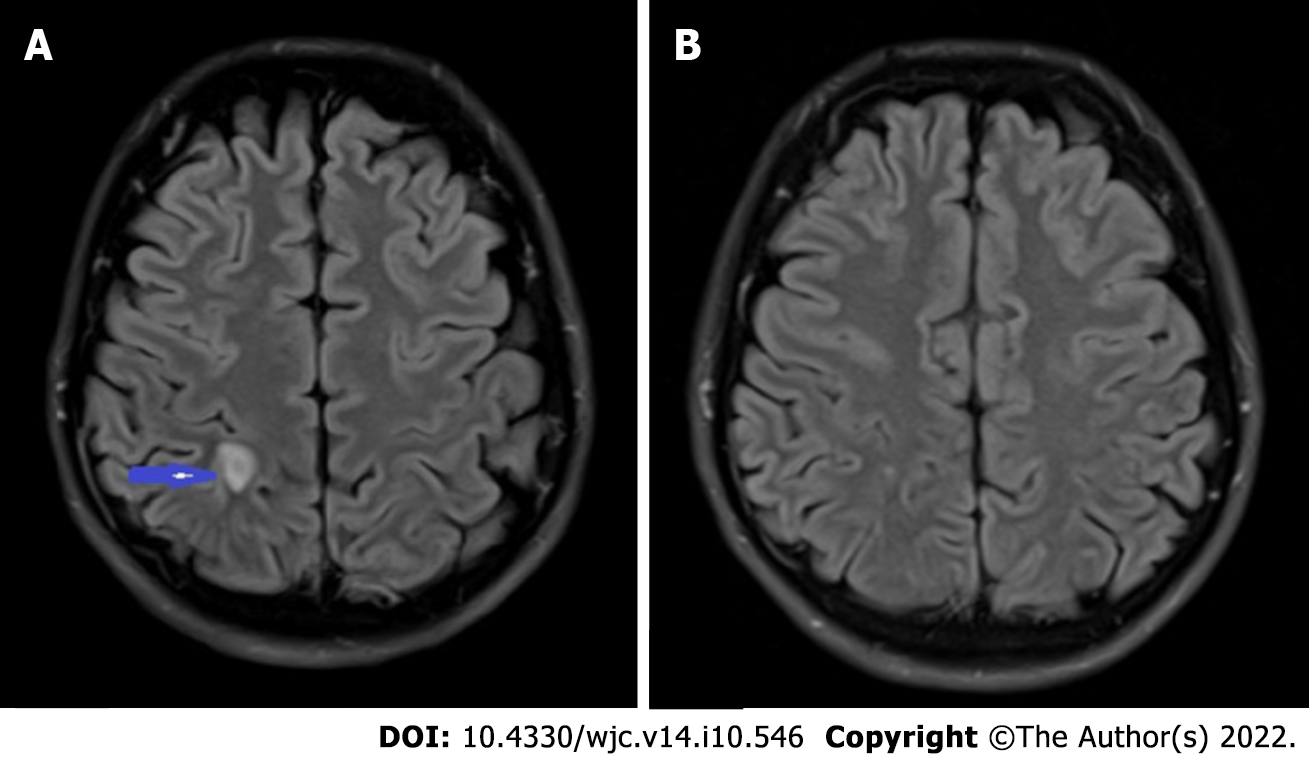
Figure 2 Septic embolus in the brain.
A: T2-weighted MRI brain showing a 1.0 cm × 0.5 cm ring enhancing lesion (blue arrow) in the right parietal lobe with central diffusion restriction and mild surrounding vasogenic edema; B: Repeat MRI brain with near complete resolution of the ring enhancing lesion. MRI: Magnetic resonance imaging.
- Citation: Olagunju A, Martinez J, Kenny D, Gideon P, Mookadam F, Unzek S. Virulent endocarditis due to Haemophilus parainfluenzae: A systematic review of the literature. World J Cardiol 2022; 14(10): 546-556
- URL: https://www.wjgnet.com/1949-8462/full/v14/i10/546.htm
- DOI: https://dx.doi.org/10.4330/wjc.v14.i10.546


