Copyright
©The Author(s) 2021.
World J Cardiol. Sep 26, 2021; 13(9): 416-437
Published online Sep 26, 2021. doi: 10.4330/wjc.v13.i9.416
Published online Sep 26, 2021. doi: 10.4330/wjc.v13.i9.416
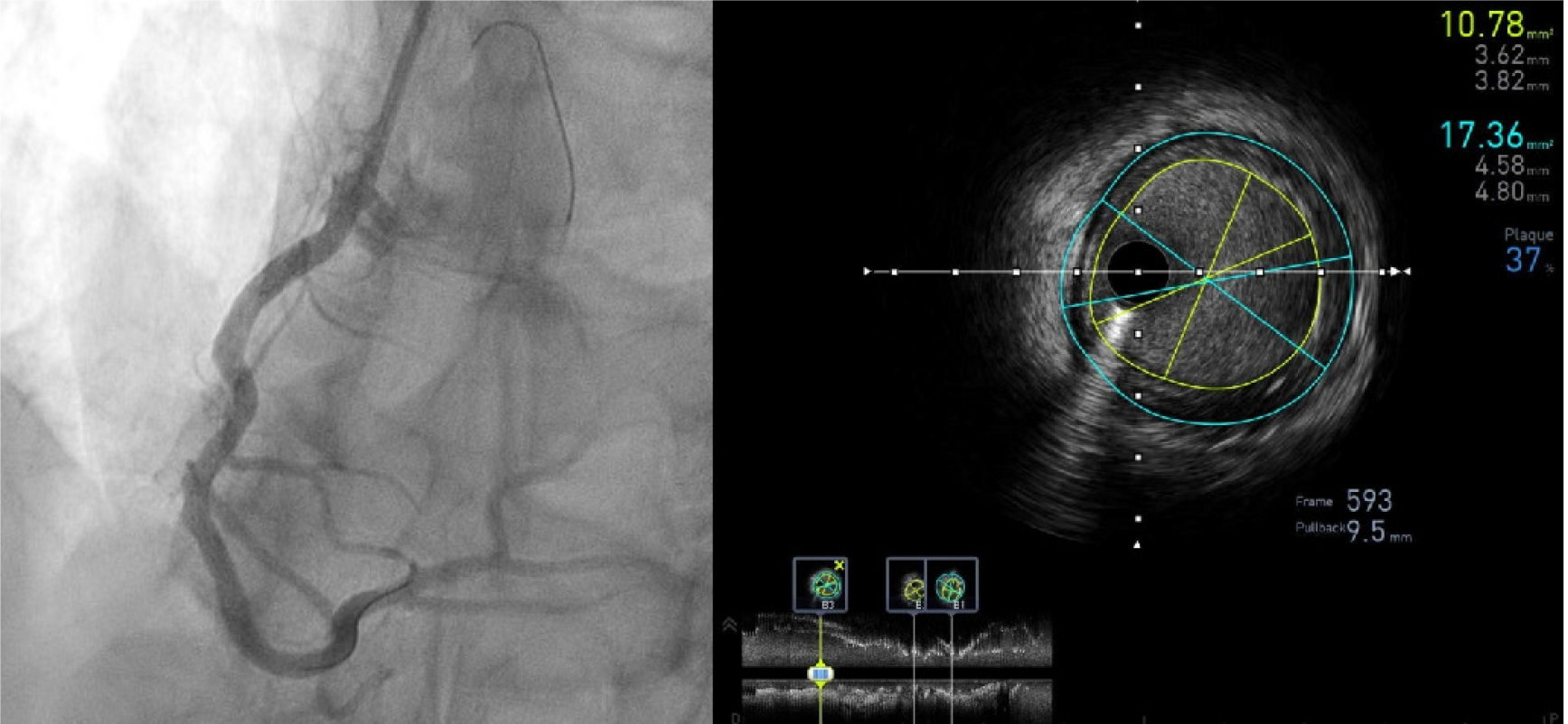
Figure 3 Intravascular ultrasound pull-back of a right coronary artery post stent implantation.
Stent expansion is assessed and the minimal lumen area measured in different cross-sections (bottom longitudinal view) while the plaque burden at the distal reference (B3) is shown on the cross sectional frame 593 (lumen area 10.78 mm², vessel area 17.36 mm², plaque burden 37%).
- Citation: Ghafari C, Carlier S. Stent visualization methods to guide percutaneous coronary interventions and assess long-term patency. World J Cardiol 2021; 13(9): 416-437
- URL: https://www.wjgnet.com/1949-8462/full/v13/i9/416.htm
- DOI: https://dx.doi.org/10.4330/wjc.v13.i9.416


