Copyright
©The Author(s) 2021.
World J Cardiol. Aug 26, 2021; 13(8): 254-270
Published online Aug 26, 2021. doi: 10.4330/wjc.v13.i8.254
Published online Aug 26, 2021. doi: 10.4330/wjc.v13.i8.254
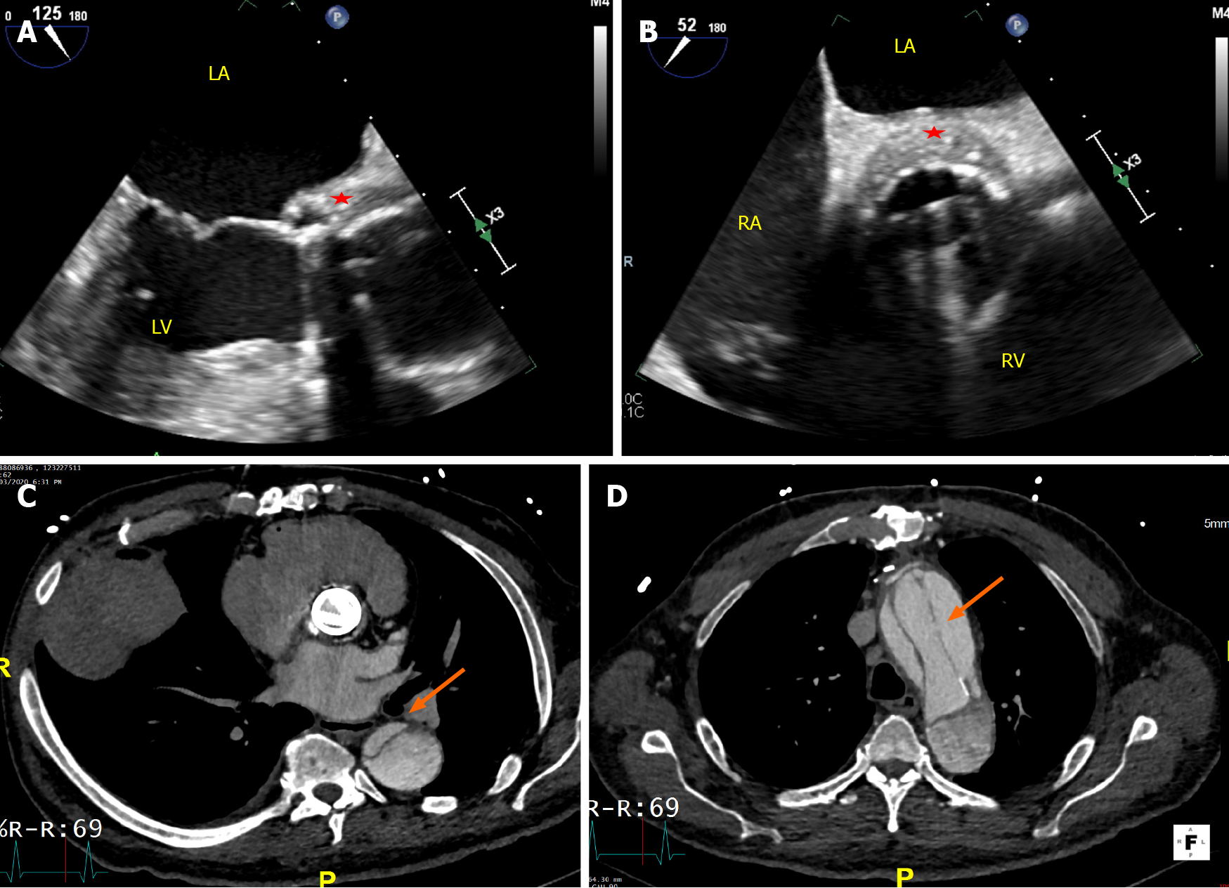
Figure 6 Transesophageal echocardiogram and cardiac computed tomography in a patient with metallic aortic valve replacement endo
- Citation: Lo Presti S, Elajami TK, Zmaili M, Reyaldeen R, Xu B. Multimodality imaging in the diagnosis and management of prosthetic valve endocarditis: A contemporary narrative review. World J Cardiol 2021; 13(8): 254-270
- URL: https://www.wjgnet.com/1949-8462/full/v13/i8/254.htm
- DOI: https://dx.doi.org/10.4330/wjc.v13.i8.254


