Copyright
©The Author(s) 2021.
World J Cardiol. Aug 26, 2021; 13(8): 254-270
Published online Aug 26, 2021. doi: 10.4330/wjc.v13.i8.254
Published online Aug 26, 2021. doi: 10.4330/wjc.v13.i8.254
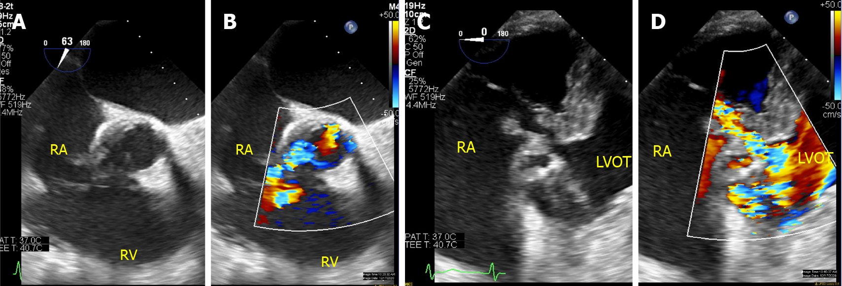
Figure 3 Para-valvular fistula in a patient with complicated endocarditis involving prior stentless aortic valve.
Eighty-two year-old male with a history of coronary artery bypass grafts and stentless aortic valve replacement presenting with Streptococcus Gallolyticus prosthetic valve endocarditis complicated by aortic root abscess and fistulous tract treated with homograft aortic root replacement (25 mm Cryo-Life aortic allograft root), bovine pericardial closure of aortic root to right atrium fistula, right atrium reconstruction with bovine patch and bypass revision. A and B: TEE interrogating the aortic valve replacement with color compare demonstrated thickening of the leaflets and abnormal color flow into the right atrium; C and D: Additional imaging from deep transgastric views with color compare in the left ventricular outflow tract demonstrated a complex fistulous tract (Gerbode-type defect) communicating the left ventricular outflow track, right atrium and aortic root. RA: Right atrium; RV: Right ventricle; LVOT: Left ventricular outflow tract.
- Citation: Lo Presti S, Elajami TK, Zmaili M, Reyaldeen R, Xu B. Multimodality imaging in the diagnosis and management of prosthetic valve endocarditis: A contemporary narrative review. World J Cardiol 2021; 13(8): 254-270
- URL: https://www.wjgnet.com/1949-8462/full/v13/i8/254.htm
- DOI: https://dx.doi.org/10.4330/wjc.v13.i8.254


