Copyright
©The Author(s) 2021.
World J Cardiol. Jul 26, 2021; 13(7): 204-210
Published online Jul 26, 2021. doi: 10.4330/wjc.v13.i7.204
Published online Jul 26, 2021. doi: 10.4330/wjc.v13.i7.204
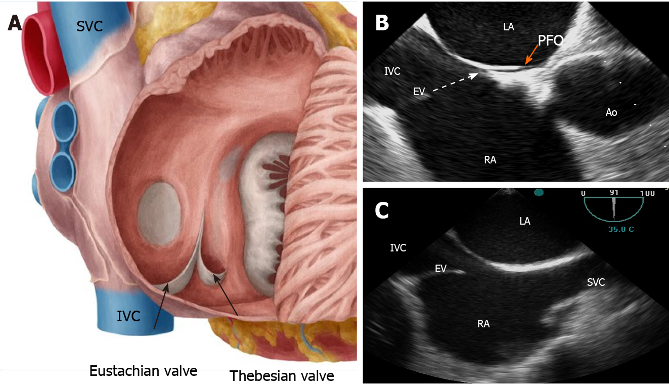
Figure 2 Anatomical illustration (A) and two-dimensional transesophageal echocardiogram in sagittal (B) and in bicaval (C) views showing the specific orientation of the eustachian valve directing the blood (white dotted arrow) toward the interatrial septum and patent foramen ovale (orange arrow).
SVC: Superior vena cava; IVC: Inferior vena cava; RA: Right atrium; LA: Left atrium; EV: Eustachian valve; Ao: Aorta; PFO: Patent foramen ovale.
- Citation: Onorato EM. Large eustachian valve fostering paradoxical thromboembolism: passive bystander or serial partner in crime? . World J Cardiol 2021; 13(7): 204-210
- URL: https://www.wjgnet.com/1949-8462/full/v13/i7/204.htm
- DOI: https://dx.doi.org/10.4330/wjc.v13.i7.204


