Copyright
©The Author(s) 2021.
World J Cardiol. Jun 26, 2021; 13(6): 155-162
Published online Jun 26, 2021. doi: 10.4330/wjc.v13.i6.155
Published online Jun 26, 2021. doi: 10.4330/wjc.v13.i6.155
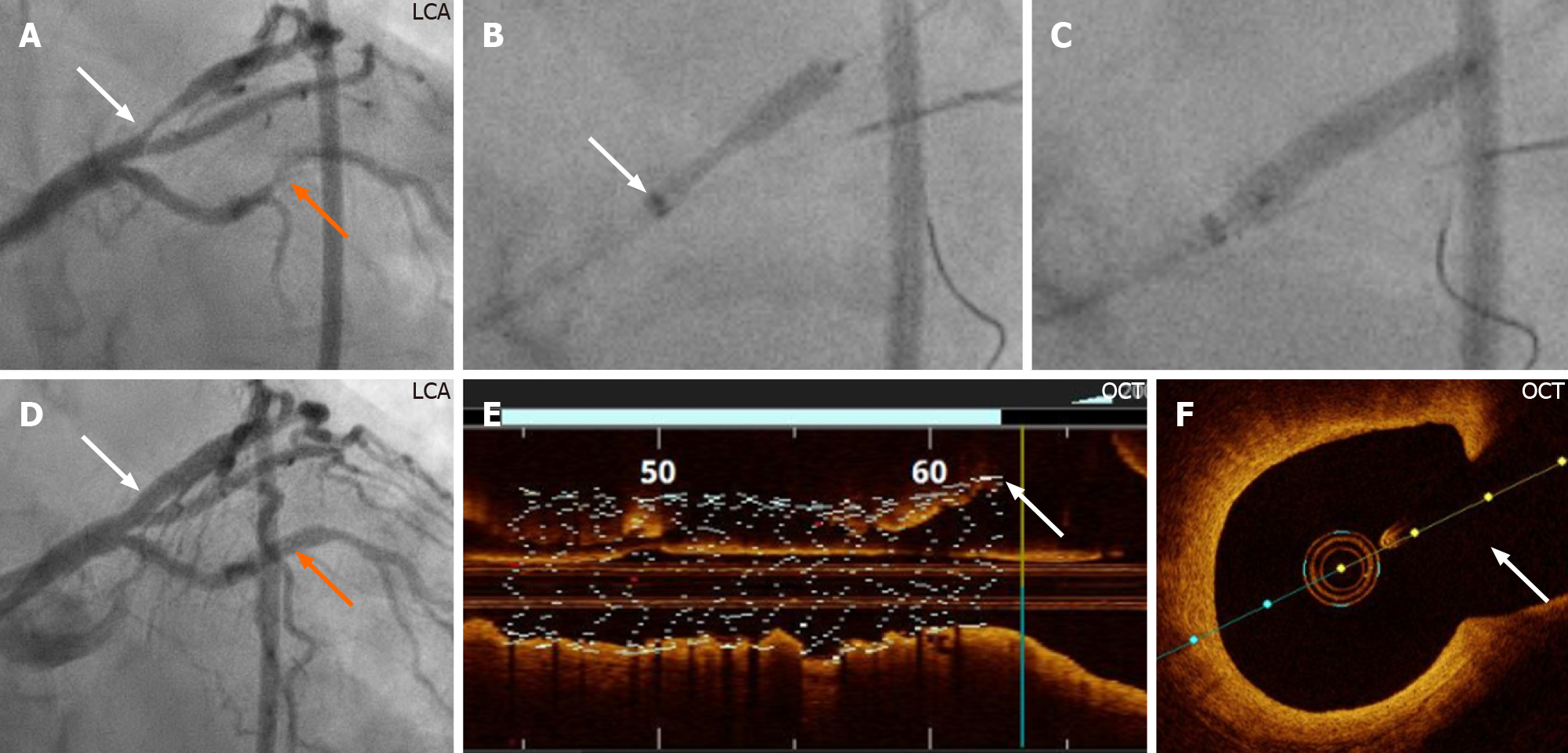
Figure 3 Demonstration of the guide extension technique during percutaneous coronary intervention of the bifurcation lesion of the second case.
A: Left coronary artery (LCA) angiography reveals a stenosis in the obtuse marginal branch, (orange arrow) that was stented first after engagement of the left coronary artery with 7F-XB 3.5 guide catheter. There is also a distal mono-ostial (medina 0, 1, 0) left anterior descending artery (LAD)/diagonal bifurcation stenosis (white arrow), which was stented with the guide extension-assisted technique; B: Of note, angiography to visualize the angle between the diagonal branch and the distal segment of the main branch (LAD) was ambiguous, making it especially suitable for this novel technique. The GuideLiner, mounted on both the LAD and diagonal guidewires, was introduced to the carina site of the bifurcation. A Promus Premier 3.0 mm × 16 mm stent (Boston Scientific, Marlborough, MA, United States) positioned at the stenosis with the proximal stent balloon radio-opaque marker positioned on the GuideLiner radio-opaque marker just proximal to the tip of the GuideLiner (white arrow); C: The stent was implanted at a pressure of 22 atm; D: Angiographic final result (white and orange arrows); E and F: The implanted stent, checked by using OCT, is well-apposed to the vessel wall; the proximal stent edge can be seen positioned at the ostium of the distal segment of the main branch (E, white arrow), and no stent struts are seen to cover the diagonal branch ostium (F, white arrow). OCT: Optical coherence tomography.
- Citation: Y-Hassan S, de Palma R. A Novel guide extension assisted stenting technique for coronary bifurcation lesions. World J Cardiol 2021; 13(6): 155-162
- URL: https://www.wjgnet.com/1949-8462/full/v13/i6/155.htm
- DOI: https://dx.doi.org/10.4330/wjc.v13.i6.155


