Copyright
©The Author(s) 2021.
World J Cardiol. Jun 26, 2021; 13(6): 155-162
Published online Jun 26, 2021. doi: 10.4330/wjc.v13.i6.155
Published online Jun 26, 2021. doi: 10.4330/wjc.v13.i6.155
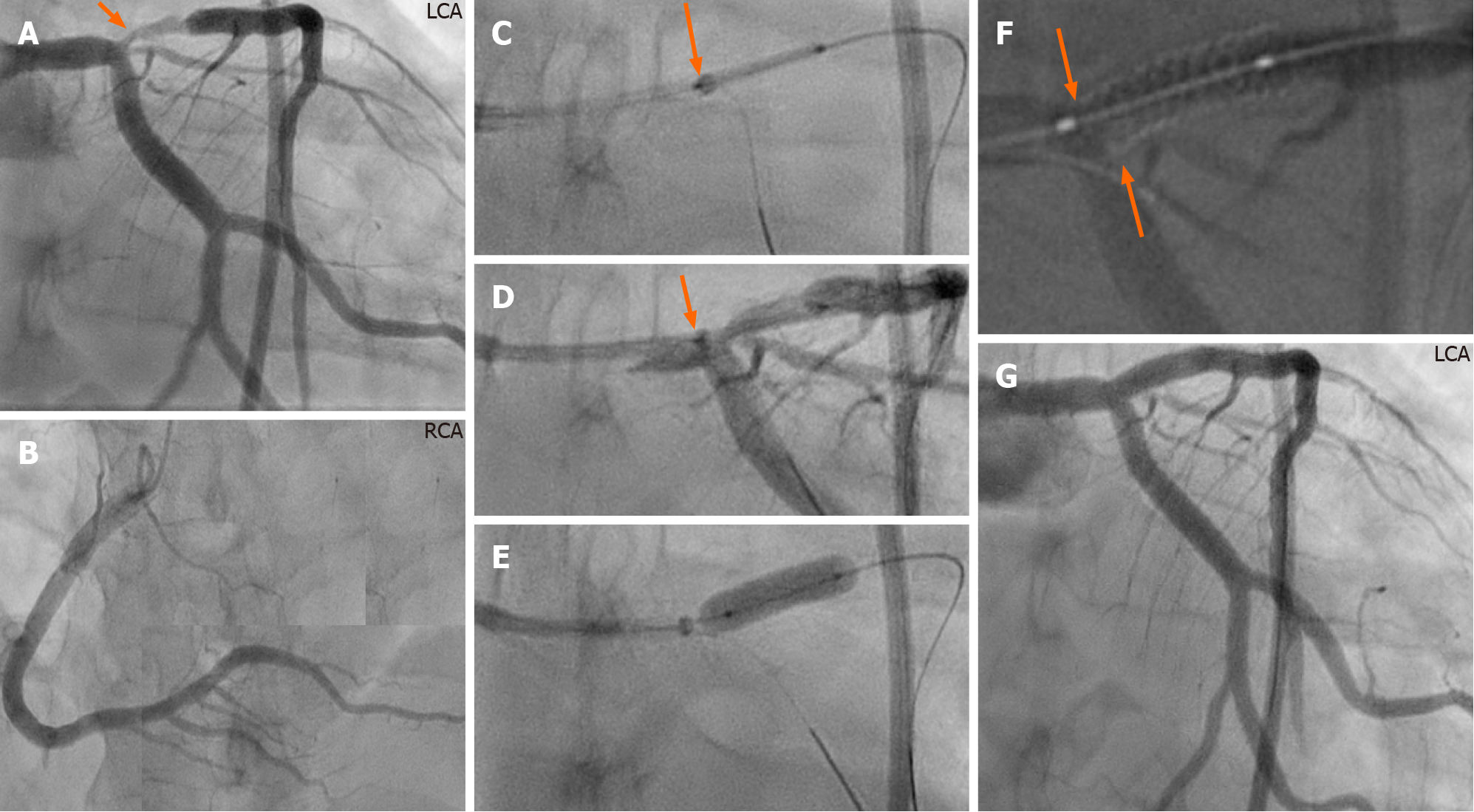
Figure 2 Annotated description of the guide extension technique in the bifurcation lesion in the first case.
A and B: Left (A) and right (B) coronary angiography of the same patient in figure 1 showing stenosis at the ostium and proximal part of left anterior descending artery (LAD) (A, orange arrow); C and D: After engagement of the left coronary artery with 7F XB 3.5 guide catheter, a 7F GuideLiner, mounted on both guidewires, is advanced carefully against the carina of the bifurcation and the stent is placed at the stenosis site with the proximal radio-opaque marker of the stent balloon overlapping the radio-opaque marker just proximal to the tip of the GuideLiner (D, under contrast injection); E: A 3.5 mm × 15 mm stent was implanted with a pressure of 20 atm; note that the GuideLiner is pushed backwards during stent implantation. The stent is post-dilated in the conventional way; F: The proximal stent edge can be seen accurately placed at the ostium using StentBoost imaging (orange arrows); G: Final result, stent implanted in the LAD without compromising the left main stem or the left circumflex artery ostium. LCA: Left coronary artery; RCA: Right coronary artery.
- Citation: Y-Hassan S, de Palma R. A Novel guide extension assisted stenting technique for coronary bifurcation lesions. World J Cardiol 2021; 13(6): 155-162
- URL: https://www.wjgnet.com/1949-8462/full/v13/i6/155.htm
- DOI: https://dx.doi.org/10.4330/wjc.v13.i6.155


