Copyright
©The Author(s) 2021.
World J Cardiol. Jun 26, 2021; 13(6): 155-162
Published online Jun 26, 2021. doi: 10.4330/wjc.v13.i6.155
Published online Jun 26, 2021. doi: 10.4330/wjc.v13.i6.155
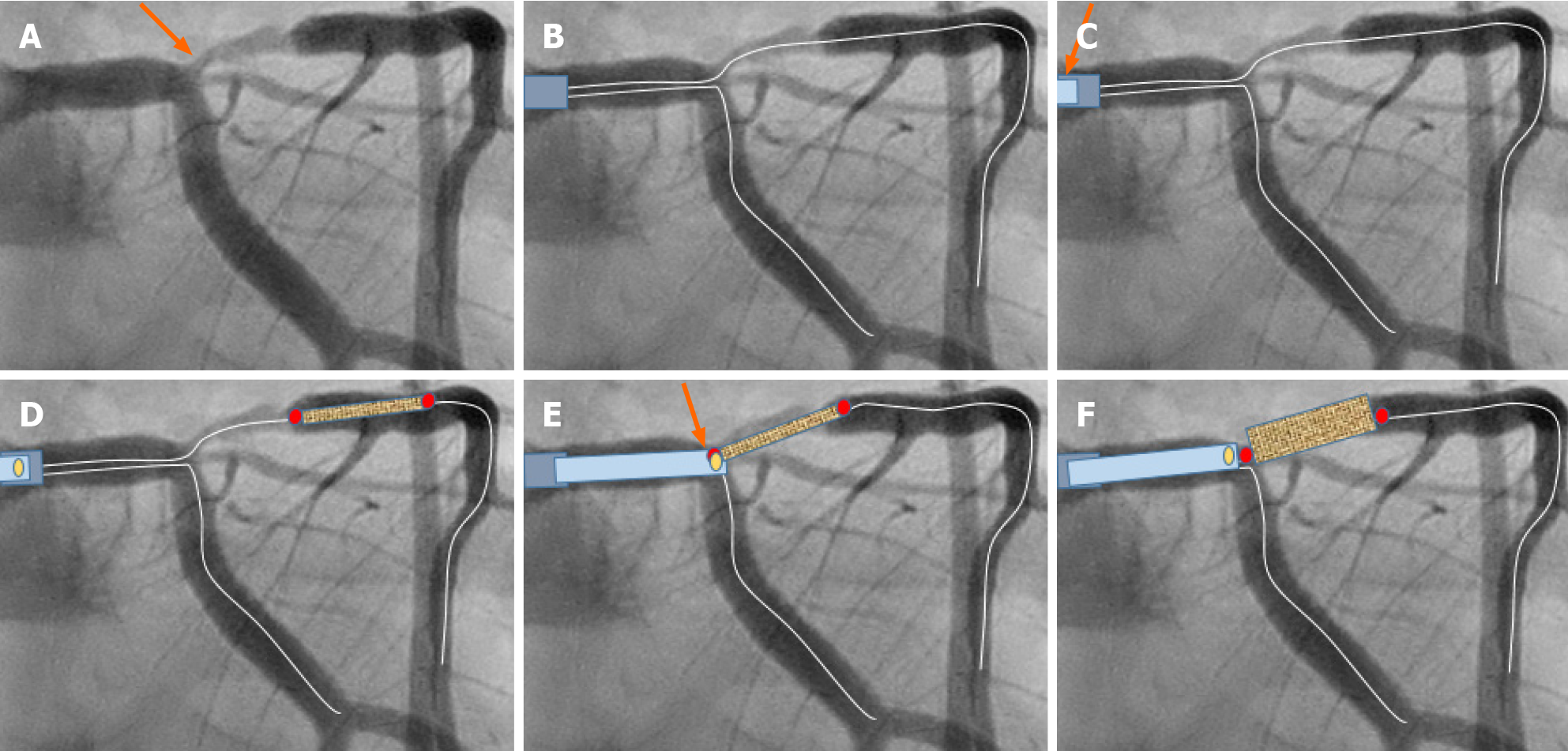
Figure 1 Schematic description of the guide extension-assisted technique in a coronary bifurcation lesion in the first case.
A: Coronary angiography in a male patient admitted with unstable angina revealed a significant ostial and proximal left anterior descending artery (LAD) stenosis. It was classified as side branch mono-ostial (medina 0, 0, 1), left main stem/left circumflex artery (LCx)/LAD bifurcation stenosis (orange arrow). Evaluation by fractional flow reserve assessment was 0.76, indicating a significant stenosis. The guide extension-assisted percutaneous coronary intervention technique is demonstrated schematically. B: Two guide wires are placed in the left coronary artery (both LAD and LCx); C: After conventional pre-dilatation, the GuideLiner, mounted on both guidewires, is placed proximal to the tip of the mother guide catheter (orange arrow); D: The stent is introduced just distal to the stenosis; E: The GuideLiner is then advanced to the carina of the bifurcation and the stent is pulled back to a level where the proximal radio-opaque marker of the stent balloon overlaps the radio-opaque marker just proximal to the tip of the guide-liner (orange arrow). Because the GuideLiner is mounted on both wires, the carina of the bifurcation will prevent further introduction of the GuideLiner into the LAD or LCx; F: The stent is implanted slowly. Note that the GuideLiner is pushed backwards when the stent balloon is inflated.
- Citation: Y-Hassan S, de Palma R. A Novel guide extension assisted stenting technique for coronary bifurcation lesions. World J Cardiol 2021; 13(6): 155-162
- URL: https://www.wjgnet.com/1949-8462/full/v13/i6/155.htm
- DOI: https://dx.doi.org/10.4330/wjc.v13.i6.155


