Copyright
©The Author(s) 2021.
World J Cardiol. Apr 26, 2021; 13(4): 111-116
Published online Apr 26, 2021. doi: 10.4330/wjc.v13.i4.111
Published online Apr 26, 2021. doi: 10.4330/wjc.v13.i4.111
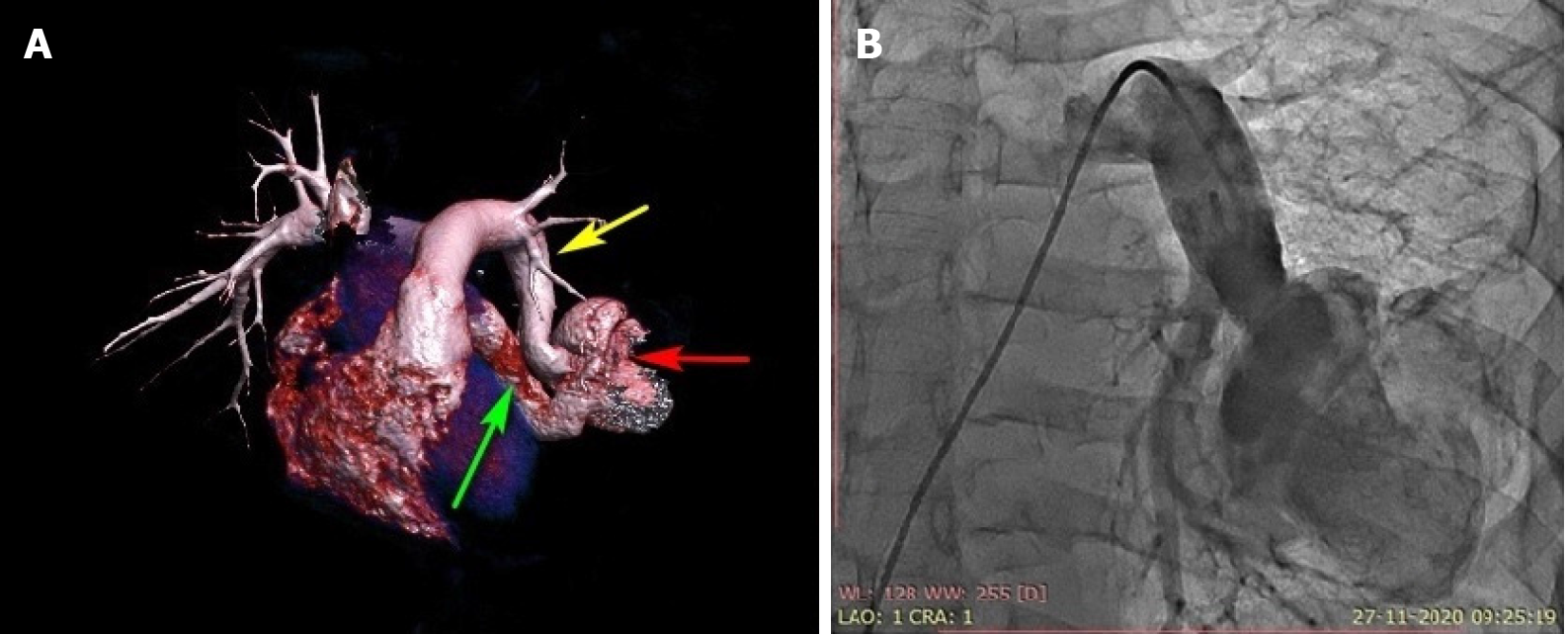
Figure 1 Three-dimensional computed tomography volume rendering and pulmonary artery angiogram.
A: Contrast-enhanced computed tomography with three-dimensional reconstruction showing the left lower pulmonary artery-to-left atrial fistula with a highly tortuous course associated with an aneurysmal sac. Yellow arrow: left lower segmental artery; red arrow: aneurysmal sac; green arrow: last part of fistula connecting the left atrium; B: Anterior-posterior projection of pulmonary artery angiography using a 6-Fr pigtail catheter showing a highly tortuous large left pulmonary artery-to-left atrial fistula associated with a 6-cm aneurysmal sac. The right ventricular oxygen saturation was 79%, and the left ventricular saturation was 92% in room air without anesthesia.
- Citation: Mahapatra R, Mahanta D, Singh J, Acharya D, Barik R. Device closure of fistula from left lower pulmonary artery to left atrium using a vascular plug: A case report. World J Cardiol 2021; 13(4): 111-116
- URL: https://www.wjgnet.com/1949-8462/full/v13/i4/111.htm
- DOI: https://dx.doi.org/10.4330/wjc.v13.i4.111


