Copyright
©The Author(s) 2021.
World J Cardiol. Nov 26, 2021; 13(11): 628-649
Published online Nov 26, 2021. doi: 10.4330/wjc.v13.i11.628
Published online Nov 26, 2021. doi: 10.4330/wjc.v13.i11.628
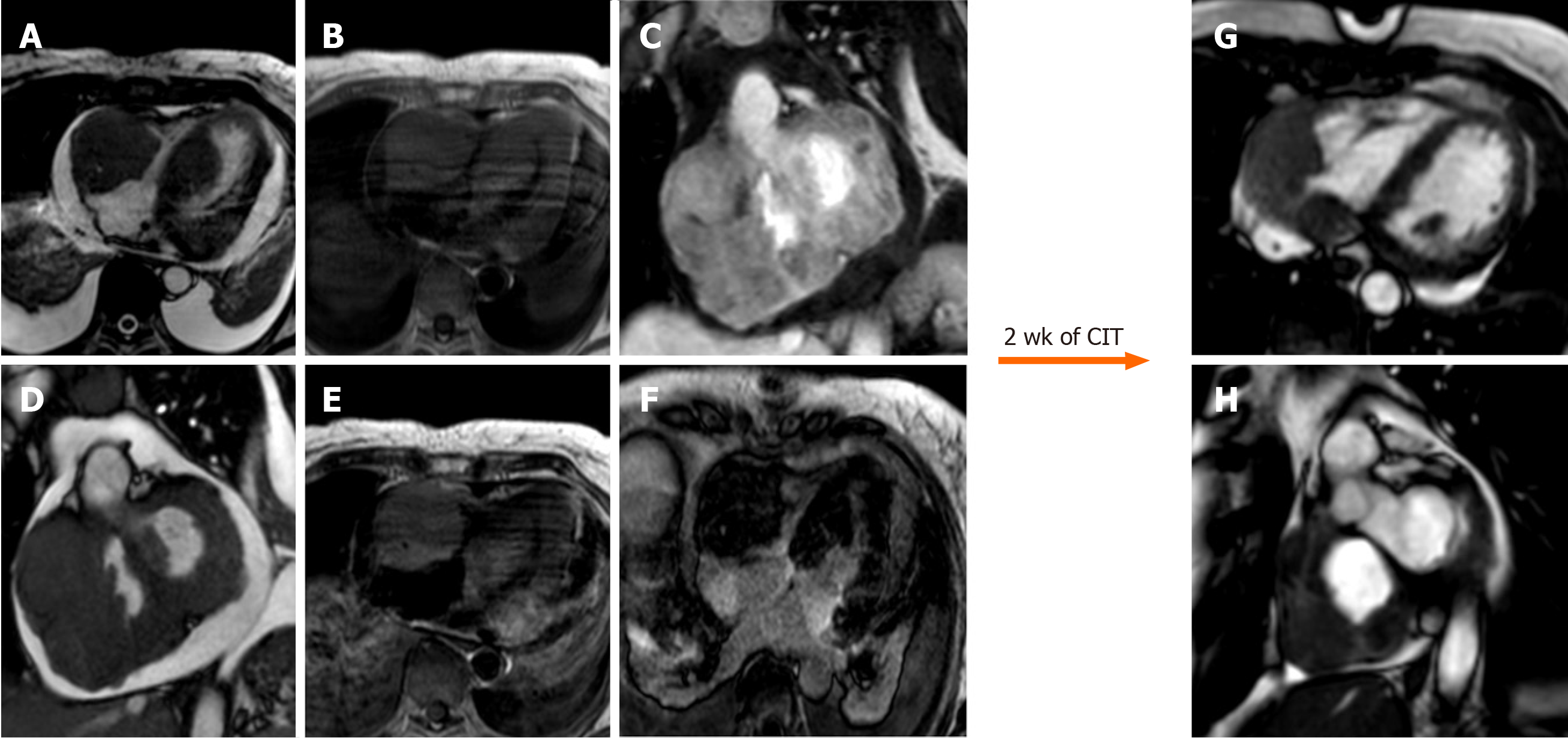
Figure 12 Sixty-five-year-old female with transthoracic echocardiogram finding of two masses in the left ventricle and right cavities following an episode of lipothymia.
Cardiovascular magnetic resonance (CMR) with cine-steady state free precession images (A and B) demonstrates the presence of four distinct masses, the most voluminous mass with infiltrative features and irregular margins, extending from the right atrio-ventricular groove to both atrial and the ventricular walls. These lesions are characterized by a substantially isointense signal to cardiac muscle in T1-weighted images (C), slightly hyperintense in T2-weighted images (D) with heterogeneous enhancement at early gadolinium enhancement and relatively homogeneous hypointensity at late gadolinium enhancement images (E, F). These findings are in keeping with cardiac lymphoma. The patient underwent biopsy with a diagnosis of cardiac large B-cell lymphoma and started a chemoimmunotherapy with an excellent response as shown on CMR (G and H, cine steady state free precession) after 2 wk. CIT: Chemo
- Citation: Gatti M, D’Angelo T, Muscogiuri G, Dell'aversana S, Andreis A, Carisio A, Darvizeh F, Tore D, Pontone G, Faletti R. Cardiovascular magnetic resonance of cardiac tumors and masses. World J Cardiol 2021; 13(11): 628-649
- URL: https://www.wjgnet.com/1949-8462/full/v13/i11/628.htm
- DOI: https://dx.doi.org/10.4330/wjc.v13.i11.628


