Copyright
©The Author(s) 2021.
World J Cardiol. Nov 26, 2021; 13(11): 628-649
Published online Nov 26, 2021. doi: 10.4330/wjc.v13.i11.628
Published online Nov 26, 2021. doi: 10.4330/wjc.v13.i11.628
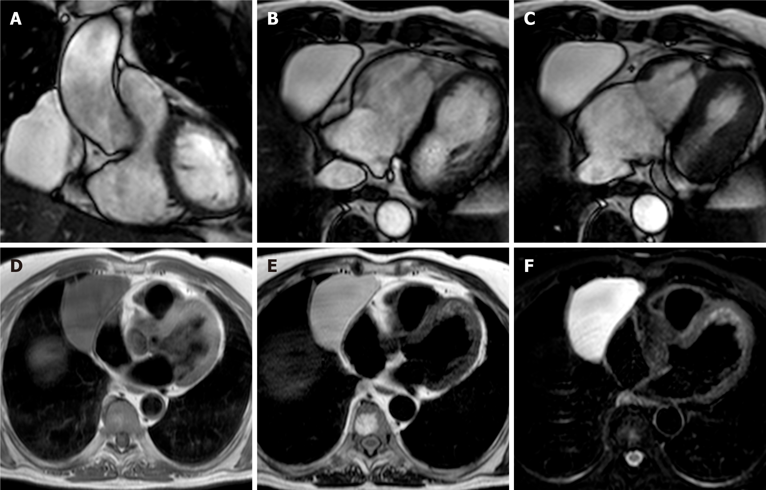
Figure 5 Fifty-seven-year-old female patient with parenchymal mass in the right cardiophrenic angle on chest X-ray.
This finding was first investigated by chest computed tomography and then by cardiovascular magnetic resonance (CMR) (A-C). The CMR confirm the presence of the mass on cine-steady state free precession images (A and B, two orthogonal diastolic frames of plane through the mass respectively), without sign of infiltration confirmed by the preserved movement of the heart chambers in relation to the mass itself. The mass shows low signal on T1-weighted sequences (D) and high signal on T2-weighted images (E and F, without and with fat suppression, respectively). These findings are in keeping with pericardial cyst.
- Citation: Gatti M, D’Angelo T, Muscogiuri G, Dell'aversana S, Andreis A, Carisio A, Darvizeh F, Tore D, Pontone G, Faletti R. Cardiovascular magnetic resonance of cardiac tumors and masses. World J Cardiol 2021; 13(11): 628-649
- URL: https://www.wjgnet.com/1949-8462/full/v13/i11/628.htm
- DOI: https://dx.doi.org/10.4330/wjc.v13.i11.628


