Copyright
©The Author(s) 2021.
World J Cardiol. Jan 26, 2021; 13(1): 21-27
Published online Jan 26, 2021. doi: 10.4330/wjc.v13.i1.21
Published online Jan 26, 2021. doi: 10.4330/wjc.v13.i1.21
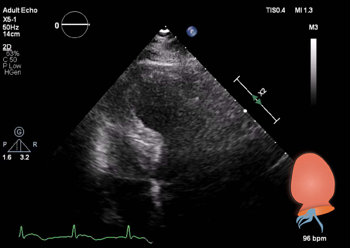
Figure 3 Transthoracic echocardiography in four-chamber view.
Apical to mid-ventricular segment ballooning was present at end-systole. Please note the endomyocardial board in end systolic contraction forming apical ballooning of the left ventricle, like a Japanese octopus trap (Takotsubo; see inset illustration), and normal right ventricle size.
- Citation: Kuo Y, Ottens TH, van der Bilt I, Keunen RW, Akin S. Myasthenic crisis-induced Takotsubo cardiomyopathy in an elderly man: A case report of an underestimated but deadly combination. World J Cardiol 2021; 13(1): 21-27
- URL: https://www.wjgnet.com/1949-8462/full/v13/i1/21.htm
- DOI: https://dx.doi.org/10.4330/wjc.v13.i1.21


