Copyright
©The Author(s) 2020.
World J Cardiol. Jun 26, 2020; 12(6): 231-247
Published online Jun 26, 2020. doi: 10.4330/wjc.v12.i6.231
Published online Jun 26, 2020. doi: 10.4330/wjc.v12.i6.231
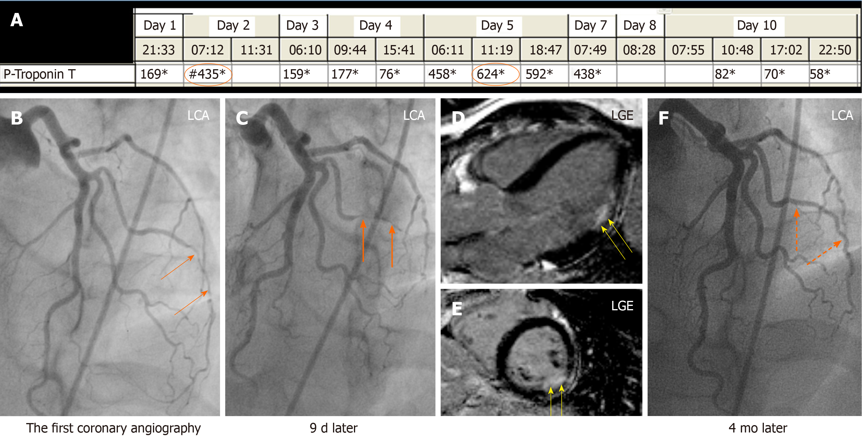
Figure 2 A case of obstructive coronary artery disease due spontaneous coronary artery dissection.
A 35-years-old female patient with a spontaneous coronary artery dissection (SCAD) of the diagonal artery with documented myocardial infarction (MI) by cardiac magnetic resonance imaging and obstructive coronary artery stenosis. A: Recurrent troponin elevation with a rise and/or fall pattern (orange circles); B: A diagonal artery with a peripheral stenosis (thin orange arrows); C: Proximal propagation of diagonal artery narrowing during repeated left coronary artery angiography 9 d later (thick orange arrows); D and E: Cardiac magnetic resonance imaging shows MI corresponding to the diagonal artery supply territory (yellow arrows); F: Follow up left coronary artery angiography 4 mo later reveals complete resolution of the diagonal artery lesion consistent with SCAD (broken orange arrows). Consequently, this patient has documented MI caused by obstructive coronary artery disease due to SCAD. LGE: Late gadolinium enhancement; LGA: Left coronary artery.
- Citation: Y-Hassan S. Autonomic neurocardiogenic syndrome is stonewalled by the universal definition of myocardial infarction. World J Cardiol 2020; 12(6): 231-247
- URL: https://www.wjgnet.com/1949-8462/full/v12/i6/231.htm
- DOI: https://dx.doi.org/10.4330/wjc.v12.i6.231


