Copyright
©The Author(s) 2020.
World J Cardiol. Feb 26, 2020; 12(2): 91-96
Published online Feb 26, 2020. doi: 10.4330/wjc.v12.i2.91
Published online Feb 26, 2020. doi: 10.4330/wjc.v12.i2.91
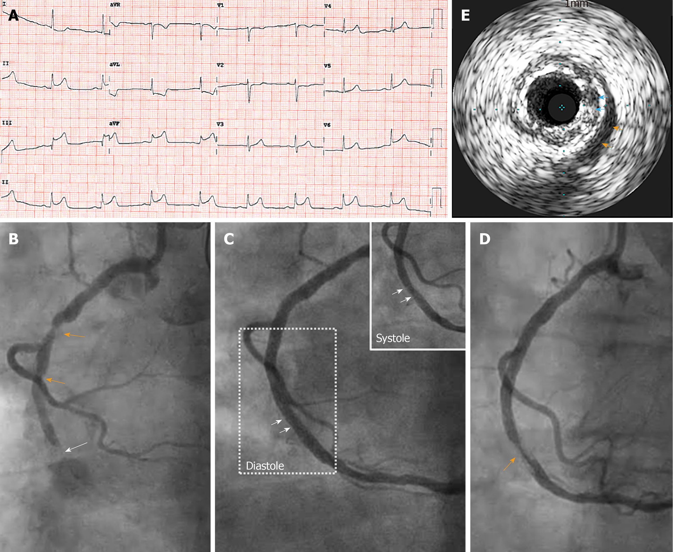
Figure 1 Images of the recurrence of inferior ST segment elevation myocardial infarction and intra-procedural findings during reperfusion therapy.
A: Presenting 12-lead electrocardiogram showed ST elevation in inferior leads; B: Culprit lesion in the mid right coronary artery (RCA) (white arrow) and diffuse atherosclerotic lesions in the proximal RCA (orange arrows); C: Initially stented RCA with systolic narrowing (insert, systolic phase of the dashed area in C) showed mild compressions of the stented lumen (white arrows); D: Circumferential filling defect in the stented segment (orange arrow); E: Intravascular ultrasound evidence of myocardial bridge with echo-lucent half-moon sign (orange arrows) and plaque between stent and myocardial bridge (blue arrows).
- Citation: Ma J, Gustafson GM, Dai X. Plaque herniation after stenting the culprit lesion with myocardial bridging in ST elevation myocardial infarction: A case report. World J Cardiol 2020; 12(2): 91-96
- URL: https://www.wjgnet.com/1949-8462/full/v12/i2/91.htm
- DOI: https://dx.doi.org/10.4330/wjc.v12.i2.91


