Copyright
©The Author(s) 2019.
World J Cardiol. Dec 26, 2019; 11(12): 316-321
Published online Dec 26, 2019. doi: 10.4330/wjc.v11.i12.316
Published online Dec 26, 2019. doi: 10.4330/wjc.v11.i12.316
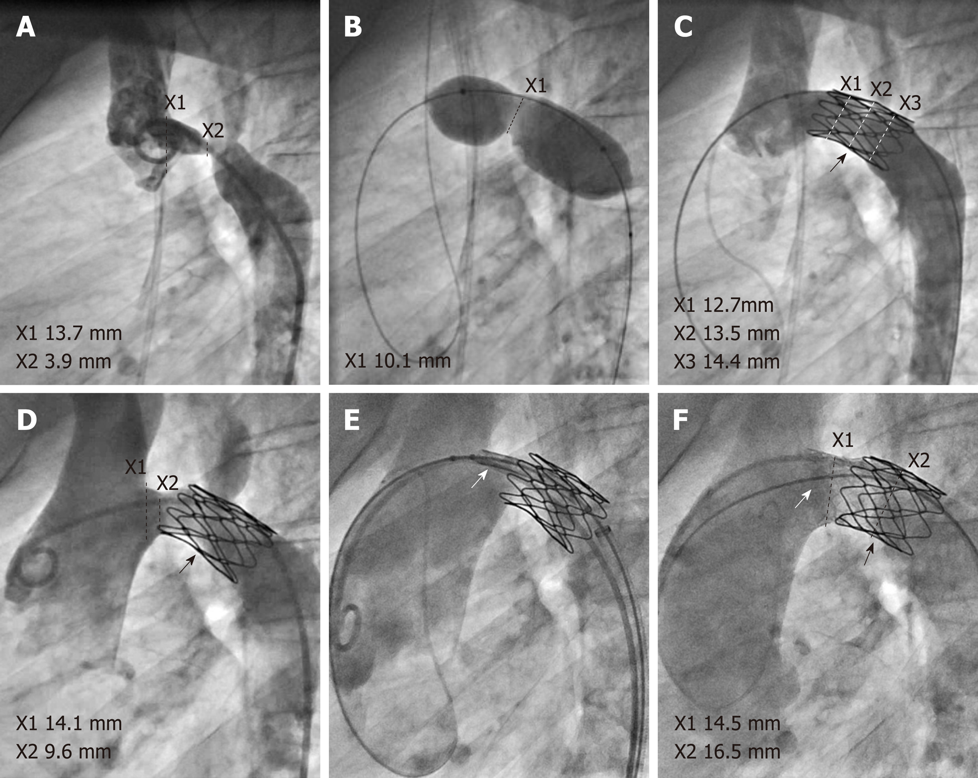
Figure 1 Angiograms in lateral projection (all images, LAO 90°) demonstrating pre-, intra- and post-interventional findings.
A: Left aortic arch with bi-carotid trunc and transverse arch hypoplasia with severe native stenosis just proximal to origin of the left subclavian artery; B: Balloon interrogation using an 18 mm Tyshak II that unmasks a relatively high compliance of the stenosis; C: After implantation of a 22 mm Cheatham-Platinum (CP) stent (indicated by black arrow) on 14 mm BiB; D: Re-stenosis proximal of the previously implanted CP stent on follow-up; E: Positioning and implantation of a LD Max 26 mm stent (indicated by white arrow) over the re-stenosis; F: Final result following redilation of both stents with 16 mm Atlas balloon, and proximal stent flaring with 20 mm Cristal balloon.
- Citation: Fürniss HE, Hummel J, Stiller B, Grohmann J. Left recurrent laryngeal nerve palsy following aortic arch stenting: A case report. World J Cardiol 2019; 11(12): 316-321
- URL: https://www.wjgnet.com/1949-8462/full/v11/i12/316.htm
- DOI: https://dx.doi.org/10.4330/wjc.v11.i12.316


