Copyright
©The Author(s) 2019.
World J Cardiol. Dec 26, 2019; 11(12): 282-291
Published online Dec 26, 2019. doi: 10.4330/wjc.v11.i12.282
Published online Dec 26, 2019. doi: 10.4330/wjc.v11.i12.282
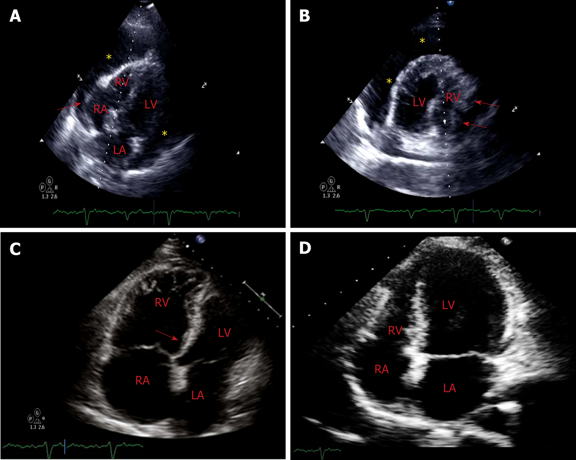
Figure 4 Computed tomography.
A: Off-axis 4-chamber view of transthoracic echocardiogram demonstrates a large circumferential pericardial effusion (marked by *) with evidence of end diastolic right chamber compression (marked by red arrow) and normal left ventricular ejection fraction. Swinging heart sign was also noted which is used to describe pendular swinging of the heart inside the pericardial space and is associated with a large pericardial effusion; B: 4-chamber view (mirror-view) demonstrates RA inversion, RV diastolic compression (marked by red arrows) and the swinging heart sign; C: Limited surface echocardiogram after pericardiocentesis demonstrated interval resolution of pericardial effusion and mild to moderately dilated right ventricle with mild RV hypokinesis and septal shift towards the left ventricle(as marked by red arrow). Patient demonstrated paradoxical worsening of blood pressure. Rapid pericardial fluid decompression may have resulted in paradoxical worsening of hemodynamics likely secondary to a combination of two factors: the first being a sudden increase in the venous return with still relatively higher systemic vascular resistance posing to a preload-afterload mismatch (hemodynamic hypothesis) and secondly likely being focal RV hypokinesis, predisposed by an increased sympathetic tone; D: A repeat echocardiogram on day 5 demonstrated normalized ventricular function. RV: Right ventricular; LV: Left ventricular; RA: Right atrial; LA: Left atrial.
- Citation: Prabhakar Y, Goyal A, Khalid N, Sharma N, Nayyar R, Spodick DH, Chhabra L. Pericardial decompression syndrome: A comprehensive review. World J Cardiol 2019; 11(12): 282-291
- URL: https://www.wjgnet.com/1949-8462/full/v11/i12/282.htm
- DOI: https://dx.doi.org/10.4330/wjc.v11.i12.282


