Copyright
©The Author(s) 2019.
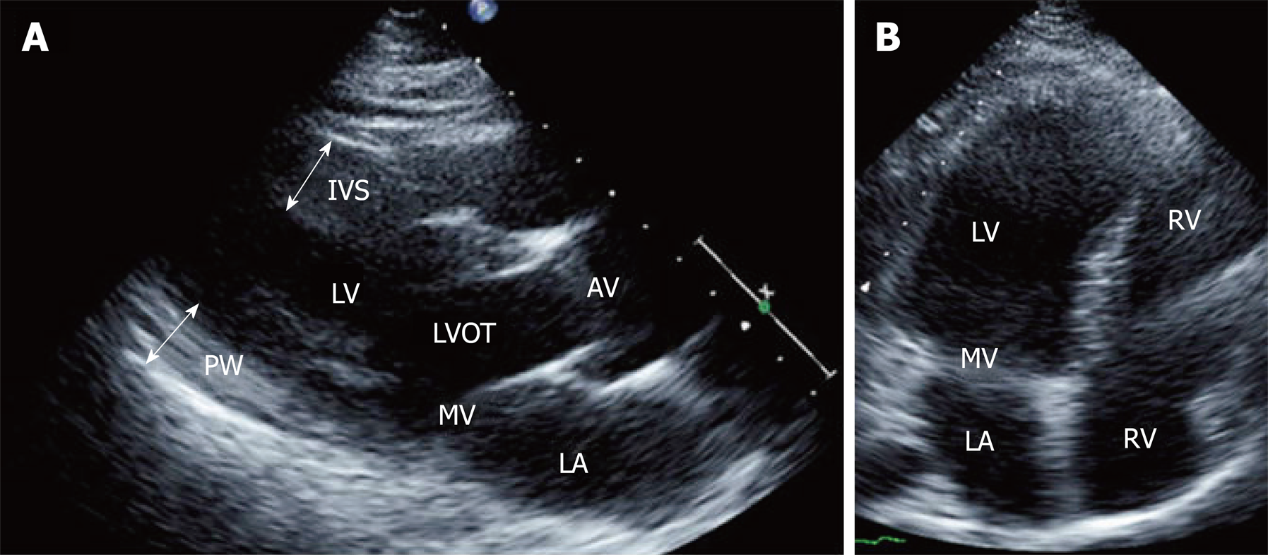
Figure 4 Two-dimensional echocardiographic still frames from a patient with Friedreich’s ataxia.
A: Parasternal long-axis view showing thickening of the interventricular septum and posterior wall; B: Apical four-chamber view showing dilatation of left ventricular cavity. IVS: Interventricular septum; PW: Posterior wall; LV: Left ventricle; LA: Left atrium; MV: Mitral valve; AV: Aortic valve; LVOT: Left ventricular outflow tract.
- Citation: Hanson E, Sheldon M, Pacheco B, Alkubeysi M, Raizada V. Heart disease in Friedreich’s ataxia. World J Cardiol 2019; 11(1): 1-12
- URL: https://www.wjgnet.com/1949-8462/full/v11/i1/1.htm
- DOI: https://dx.doi.org/10.4330/wjc.v11.i1.1


