Copyright
©The Author(s) 2019.
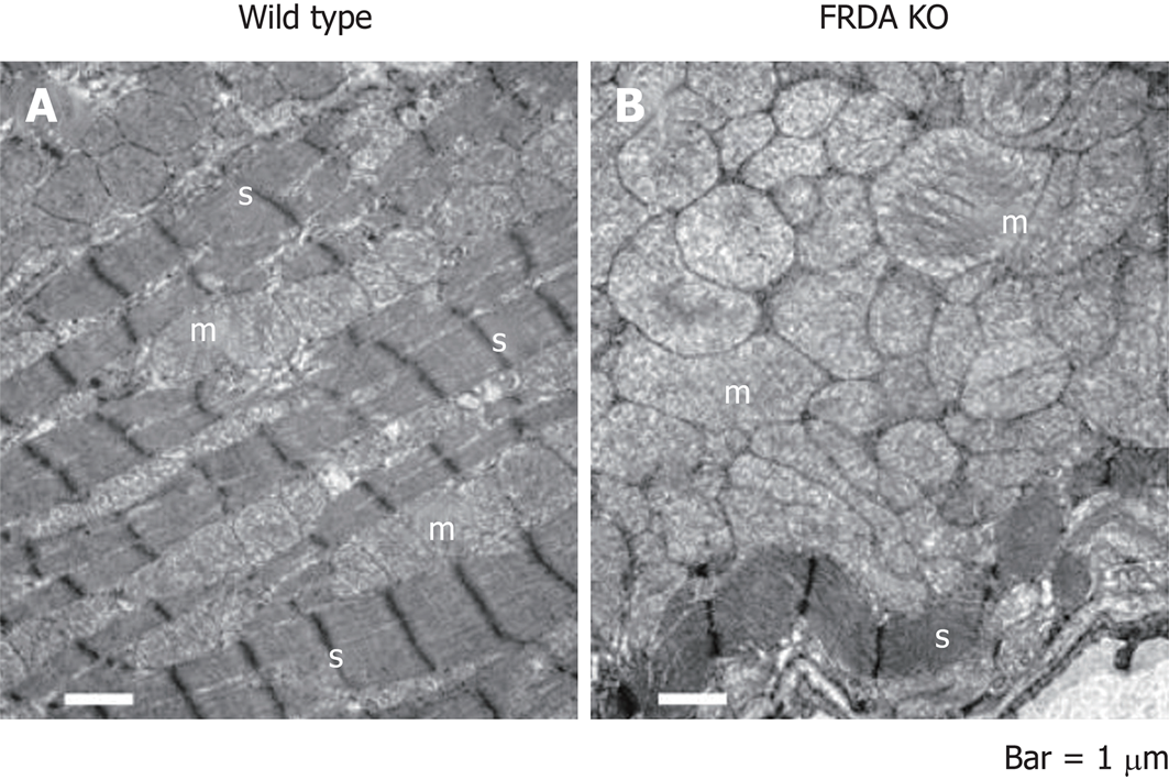
Figure 2 Electron microscopy of Friedreich’s ataxia-knockout and wild-type control mouse heart tissues from 28-d-old littermates.
A: Wild type mouse showing normal mitochondria (“m”) in rows between abundant, well-ordered sarcomeres (“s”); B: Conditional Friedreich’s ataxia-knockout (FRDA KO) mouse with ablation of the FRDA locus in the heart and brain (NSE-Cre promoter). Note the extreme proliferation of enlarged mitochondria in B. There is a severe loss of sarcomeres (“s”). Bars = 1000 nm.
- Citation: Hanson E, Sheldon M, Pacheco B, Alkubeysi M, Raizada V. Heart disease in Friedreich’s ataxia. World J Cardiol 2019; 11(1): 1-12
- URL: https://www.wjgnet.com/1949-8462/full/v11/i1/1.htm
- DOI: https://dx.doi.org/10.4330/wjc.v11.i1.1


