Copyright
©The Author(s) 2018.
World J Cardiol. Sep 26, 2018; 10(9): 110-118
Published online Sep 26, 2018. doi: 10.4330/wjc.v10.i9.110
Published online Sep 26, 2018. doi: 10.4330/wjc.v10.i9.110
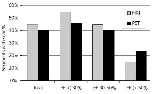
Figure 1 Histogram showing the frequency of scar detection in cardiac magnetic resonance imaging (grey bars) and positron emission tomography (black bars).
In total, cardiac magnetic resonance imaging (CMR) found scars in 45% of all segments compared to PET in 40%. CMR depicted significantly more scars in patients with severely (EF < 30%) and moderately (EF, 30%-50%) impaired left ventricular function. However, PET suggested more scars in EF > 50% group. PET: Positron emission tomography; EF: Ejection fraction.
- Citation: Hunold P, Jakob H, Erbel R, Barkhausen J, Heilmaier C. Accuracy of myocardial viability imaging by cardiac MRI and PET depending on left ventricular function. World J Cardiol 2018; 10(9): 110-118
- URL: https://www.wjgnet.com/1949-8462/full/v10/i9/110.htm
- DOI: https://dx.doi.org/10.4330/wjc.v10.i9.110


