Copyright
©The Author(s) 2018.
World J Cardiol. Nov 26, 2018; 10(11): 191-195
Published online Nov 26, 2018. doi: 10.4330/wjc.v10.i11.191
Published online Nov 26, 2018. doi: 10.4330/wjc.v10.i11.191
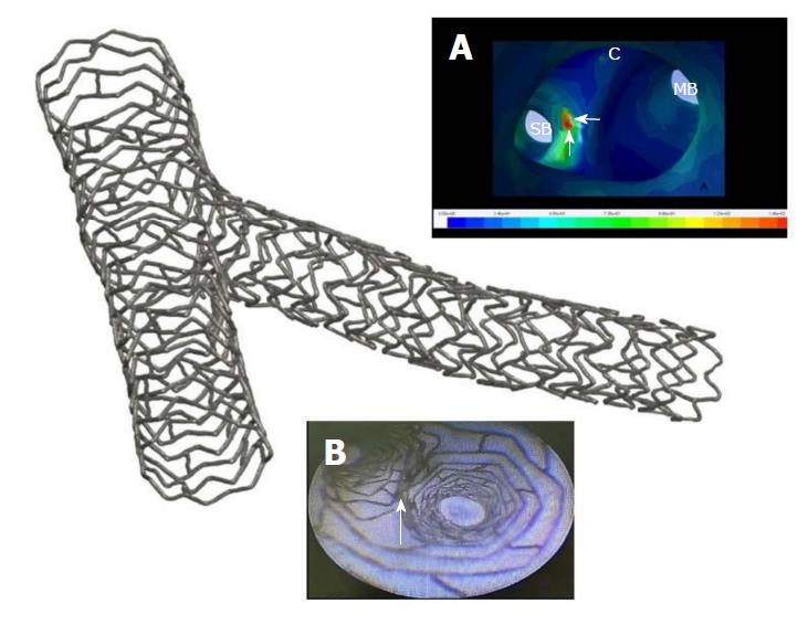
Figure 2 Microcomputed tomography picture of a bifurcation treated by the Nano-Crush technique.
A: Region of the carina investigated by computed fluid dynamic showing from the inside of a vessel with high wall shear stress (red zone, white arrows) located at the side branch portion of the carina, which should potentially be in favor of less restenosis and thrombosis at that site; B: Angioscopic image of the same region showing a very smooth transition of the wall at the bifurcation with a very minimal (Nano) apposition of two stent layers. SB: Side branch; MB: Main branch.
- Citation: Rigatelli G, Zuin M, Dash D. Thin and crush: The new mantra in left main stenting? World J Cardiol 2018; 10(11): 191-195
- URL: https://www.wjgnet.com/1949-8462/full/v10/i11/191.htm
- DOI: https://dx.doi.org/10.4330/wjc.v10.i11.191


