Copyright
©The Author(s) 2018.
World J Cardiol. Oct 26, 2018; 10(10): 165-186
Published online Oct 26, 2018. doi: 10.4330/wjc.v10.i10.165
Published online Oct 26, 2018. doi: 10.4330/wjc.v10.i10.165
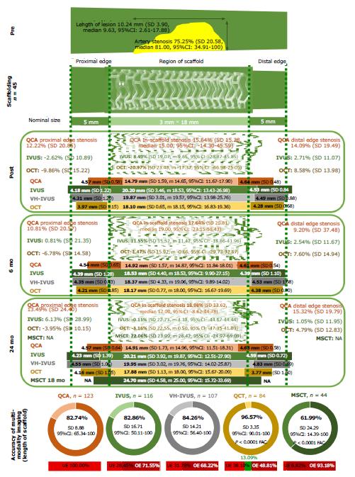
Figure 3 The accuracy of multimodality imaging analysis.
The analysis of accuracy (trueness and precision) administered with quantitative coronary angiography, intravascular ultrasound, virtual histology-IVUS, multislice computed tomography, and optical coherence tomography by the nominal length of the scaffold, which was 18 mm in all cases. The panel defines the spread-out-vessel graphics (axial resolution of 200 μm) with the appearance of the scaffolded and edge regions pre- and post-procedure at 6 mo and 24 mo. The figure was adapted from ref. [34]. UE: Underestimated (observations with the length of the scaffolded region less than 18 mm); OE: overestimated (the examined scaffolded region was more than 18 mm). QCA: quantitative coronary angiography; IVUS: intravascular ultrasound; VH-IVUS: virtual histology-intravascular ultrasound; OCT: optical coherence tomography; MSCT: multislice computed tomography.
- Citation: Kharlamov AN. Undiscovered pathology of transient scaffolding t1remains a driver of failures in clinical trials. World J Cardiol 2018; 10(10): 165-186
- URL: https://www.wjgnet.com/1949-8462/full/v10/i10/165.htm
- DOI: https://dx.doi.org/10.4330/wjc.v10.i10.165


