Copyright
©The Author(s) 2018.
World J Cardiol. Oct 26, 2018; 10(10): 153-164
Published online Oct 26, 2018. doi: 10.4330/wjc.v10.i10.153
Published online Oct 26, 2018. doi: 10.4330/wjc.v10.i10.153
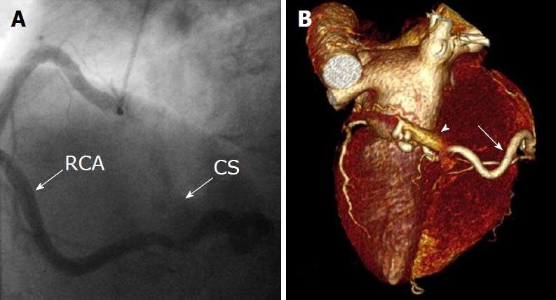
Figure 6 Unilateral fistula and computed tomography coronary angiography.
A: Unilateral fistula originating from the right coronary artery (RCA) terminating into the coronary sinus (CS) with single origin, pathway and termination with dilated RCA and enlarged CS; B: Computed tomography coronary angiography: Coronary artery fistula originating from the distal segment of RCA (arrow) and terminating into the coronary sinus. Volume-rendered three-dimensional image reconstruction demonstrating fistulous vessel located posterior connected with the CS (arrowhead). RAC: Right coronary artery; CS: Coronary sinus.
- Citation: Said SA, Agool A, Moons AH, Basalus MW, Wagenaar NR, Nijhuis RL, Schroeder-Tanka JM, Slart RH. Incidental congenital coronary artery vascular fistulas in adults: Evaluation with adenosine-13N-ammonia PET-CT. World J Cardiol 2018; 10(10): 153-164
- URL: https://www.wjgnet.com/1949-8462/full/v10/i10/153.htm
- DOI: https://dx.doi.org/10.4330/wjc.v10.i10.153


