Copyright
©The Author(s) 2024.
World J Cardiol. Feb 26, 2024; 16(2): 92-97
Published online Feb 26, 2024. doi: 10.4330/wjc.v16.i2.92
Published online Feb 26, 2024. doi: 10.4330/wjc.v16.i2.92
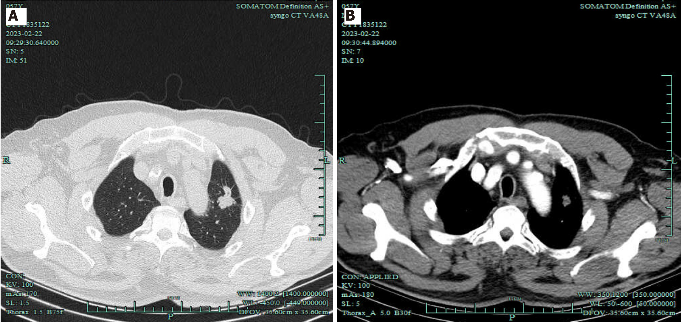
Figure 1 Imaging of pulmonary masses.
A: Chest computed tomography (CT); B: Chest CT angiography revealed 50 HU.
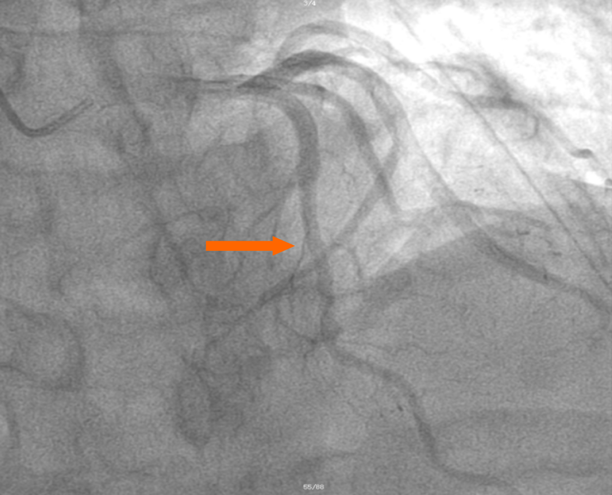
Figure 2 Partial views of coronary angiography were performed after hemostasis completion, and no abnormalities were observed in the coronary arteries (arrow).
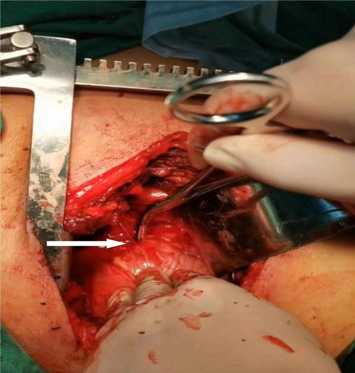
Figure 3 View of the operative field.
After opening, we revealed a bleeding point in the left anterior descending coronary artery (arrow).
- Citation: Ruan YD, Han JW. Spontaneous coronary artery rupture after lung cancer surgery: A case report and review of literature. World J Cardiol 2024; 16(2): 92-97
- URL: https://www.wjgnet.com/1949-8462/full/v16/i2/92.htm
- DOI: https://dx.doi.org/10.4330/wjc.v16.i2.92


