Copyright
©The Author(s) 2017.
World J Biol Chem. Nov 26, 2017; 8(4): 175-186
Published online Nov 26, 2017. doi: 10.4331/wjbc.v8.i4.175
Published online Nov 26, 2017. doi: 10.4331/wjbc.v8.i4.175
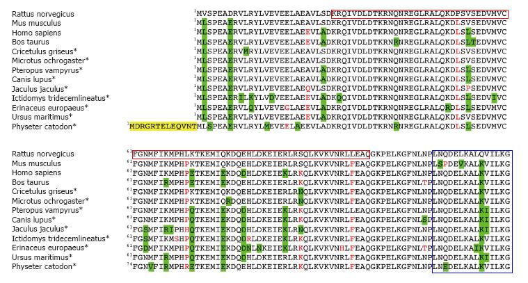
Figure 4 Alignment of mammalian PDRG1 proteins.
The figure shows the BLASTP alignment of thirteen representative mammalian PDRG1 sequences using that of Rattus norvegicus as a reference. Most of the data correspond to predicted sequences (*). Conservative changes are highlighted in green, non-conservative substitutions appear in red, and N-terminal extensions are indicated in yellow. The C-terminal helix-turn-helix motif and the prefoldin-like domain are indicated in blue and crimson boxes, respectively.
- Citation: Pajares MÁ. PDRG1 at the interface between intermediary metabolism and oncogenesis. World J Biol Chem 2017; 8(4): 175-186
- URL: https://www.wjgnet.com/1949-8454/full/v8/i4/175.htm
- DOI: https://dx.doi.org/10.4331/wjbc.v8.i4.175


