Copyright
©The Author(s) 2017.
World J Biol Chem. Aug 26, 2017; 8(3): 163-174
Published online Aug 26, 2017. doi: 10.4331/wjbc.v8.i3.163
Published online Aug 26, 2017. doi: 10.4331/wjbc.v8.i3.163
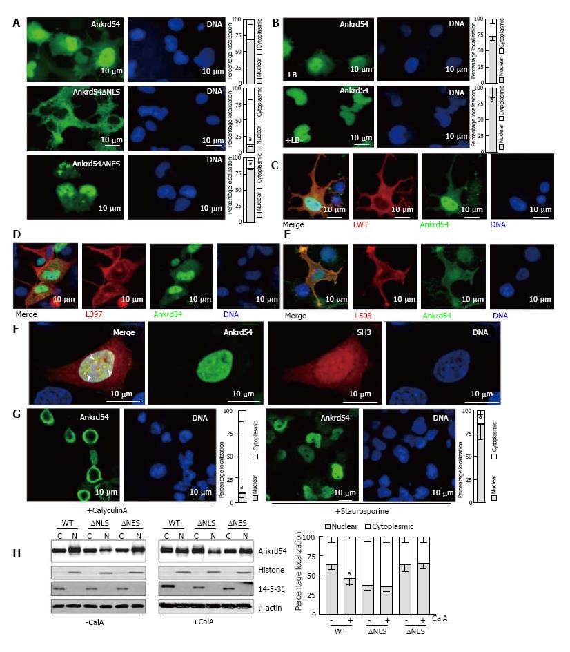
Figure 1 Ankrd54 shuttles between nuclear and cytoplasmic compartments and phosphorylation regulates this subcellular compartmentalization.
A: Localization analysis of eGFP-tagged Ankrd54 (top panel), eGFP-Ankrd54 with the NLS deleted (Ankrd54∆NLS, middle panel), and eGFP-Ankrd54 with the NES deleted (Ankrd54∆NES, bottom panel) in HEK293 cells. Delineation of the nucleus by Hoechst staining of the DNA is on the right, eGFP fluorescence is on the left. Nuclear and cytoplasmic localization of Ankrd54 was enumerated (graph at right), aP < 0.05; B: Wild-type eGFP-Ankrd54 expressing HEK293 cells were analysed after treatment with Leptomycin B (+LB) for 2 h (0.4 ng/mL), eGFP fluorescence (left) DNA counter staining (right), and quantitation of nuclear/cytoplasmic localization (graph at right), aP < 0.05; C: Localization analysis of co-expressed eGFP-Ankrd54 (green) and Lyn wild-type (LWT, red), with DNA counterstained (blue). Merged and individual channels are shown; D: Localization analysis of co-expressed eGFP-Ankrd54 (green) and kinase inactive Lyn (L397, red), with DNA counterstained (blue). Merged and individual channels are shown; E: Localization analysis of co-expressed eGFP-Ankrd54 (green) and dominant active Lyn (L598, red), with DNA counterstained (blue). Merged and individual channels are shown; F: Localization analysis of eGFP-Ankrd54 and an mCherry-fusion of the SH3 domain of Lyn, with DNA counterstained. Merged image (left panel) illustrates co-localizing nuclear puncta (arrow heads); G: Effect of CalyculinA (50 nmol/L, 60 min) or Staurosporine (100 nmol/L, 60 min) on eGFP-Ankrd54 subcellular localization. DNA counterstained (blue), eGFP fluorescence (green). Quantitation of nuclear/cytoplasmic localization (graph at right), aP < 0.05; H: Immunoblot of subcellular fractionation analysis of eGFP-Ankrd54 (WT), eGFP-Ankrd54∆NLS (∆NLS) and eGFP-Ankrd54∆NES (∆NES), without (left) and with (right) the addition of CalyculinA (50 nmol/L, 60min). Nuclear (N) and cytoplasmic (C) fractions of transfected HEK293 cells were immunoblotted using anti-Ankrd54, anti-Histone (H3), anti-14-3-3ζ, and anti-β-actin antibodies. Quantitation of nuclear/cytoplasmic localization of Ankrd54 bands depicted in graph at right, aP < 0.05.
- Citation: Samuels AL, Louw A, Zareie R, Ingley E. Control of nuclear-cytoplasmic shuttling of Ankrd54 by PKCδ. World J Biol Chem 2017; 8(3): 163-174
- URL: https://www.wjgnet.com/1949-8454/full/v8/i3/163.htm
- DOI: https://dx.doi.org/10.4331/wjbc.v8.i3.163


