Copyright
©The Author(s) 2015.
World J Biol Chem. Nov 26, 2015; 6(4): 358-365
Published online Nov 26, 2015. doi: 10.4331/wjbc.v6.i4.358
Published online Nov 26, 2015. doi: 10.4331/wjbc.v6.i4.358
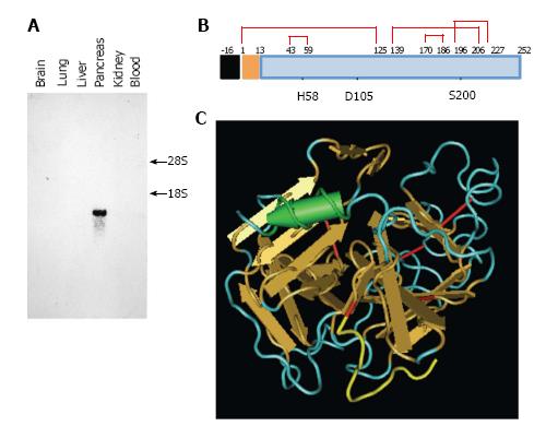
Figure 2 Caldecrin expression and protein structure.
A: Caldecrin expression was analyzed by Northern blot. 18S, 28S: 18S, 28S ribosomal RNA; B: Domain structures of caldecrin. Black box: signal peptide; orange box: pro-peptide; blue box: mature protein; red line: disulfide bridges with cysteine number; the H (histidine), D (aspartic acid), S (serine) catalytic triad; C: Ribbon diagram of the crystal structure of human caldecrin (adapted from PDB ID: 4H4F, prepared from [16]). Red line: Disulfide bridge; Yellow line: Pro-peptide; Arrow: β-sheet structure; Cylinder: α-helix structure.
- Citation: Tomomura M, Tomomura A. Caldecrin: A pancreas-derived hypocalcemic factor, regulates osteoclast formation and function. World J Biol Chem 2015; 6(4): 358-365
- URL: https://www.wjgnet.com/1949-8454/full/v6/i4/358.htm
- DOI: https://dx.doi.org/10.4331/wjbc.v6.i4.358


