Copyright
©The Author(s) 2015.
World J Biol Chem. Aug 26, 2015; 6(3): 110-120
Published online Aug 26, 2015. doi: 10.4331/wjbc.v6.i3.110
Published online Aug 26, 2015. doi: 10.4331/wjbc.v6.i3.110
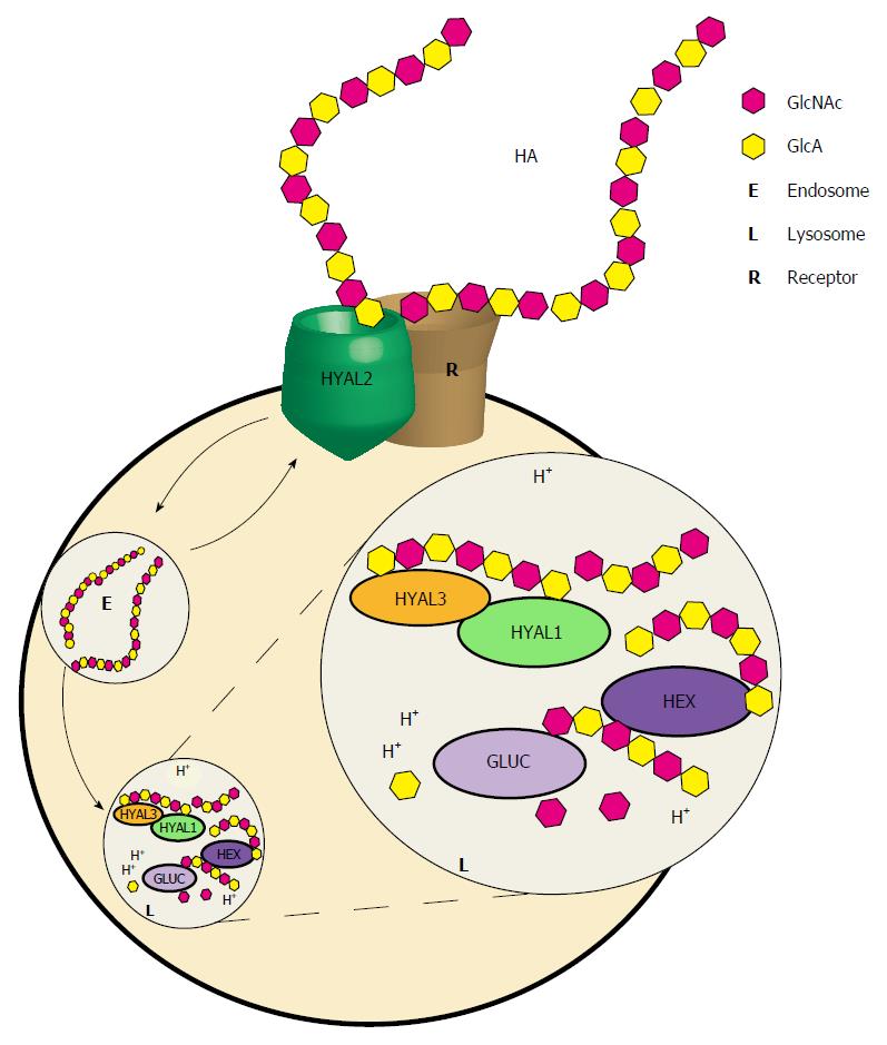
Figure 3 Proposed model for hyaluronan breakdown.
Hyaluronan (HA) bound by a cell- or matrix-associated receptor such as CD44, HARE, or LYVE-1 is proposed to be hydrolyzed to intermediate-sized fragments by the GPI-linked HYAL2. The resulting fragments are then internalized by receptor-mediated endocytosis and transported to lysosomes. Once inside the lysosome, further degradation takes place through the action of the acid-active HYAL1. HYAL1 cleaves the intermediate-sized HA fragments to smaller fragments such that they become substrates for the sequential action of the exoglycosidases, β-glucuronidase (Gluc) and β-N-acetylhexosaminidase (Hex) which hydrolyze terminal GlcA and GlcNAc, respectively. The role of HYAL3 is unclear although its overexpression increases HYAL1 activity in cell culture based studies.
- Citation: Triggs-Raine B, Natowicz MR. Biology of hyaluronan: Insights from genetic disorders of hyaluronan metabolism. World J Biol Chem 2015; 6(3): 110-120
- URL: https://www.wjgnet.com/1949-8454/full/v6/i3/110.htm
- DOI: https://dx.doi.org/10.4331/wjbc.v6.i3.110


