Copyright
©2014 Baishideng Publishing Group Inc.
World J Biol Chem. Nov 26, 2014; 5(4): 457-464
Published online Nov 26, 2014. doi: 10.4331/wjbc.v5.i4.457
Published online Nov 26, 2014. doi: 10.4331/wjbc.v5.i4.457
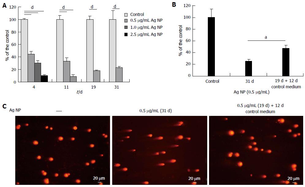
Figure 3 Low concentrations of silver nanoparticles reversibly retard human dermal microvascular endothelial cells growth.
A: Human dermal microvascular endothelial cells (HMEC) were exposed to various concentrations of silver nanoparticles (Ag NP). All the samples were trypsinized and counted on the same day in which the controls reached confluence. After counting, the cells were re-seeded at the same density; B: On day 19, part of the samples were seeded in the absence of Ag NP and counted at day 31. In (A) and (B) data are expressed as the percentage of control. Data represent the means ± SD of four separate experiments. In (A) P value was calculated vs untreated cells, dP < 0.001; and in (B) P value was calculated vs cells cultured for 31 d continuously with Ag NP, aP < 0.05; C: Comet assay was performed as described on HMEC treated with Ag NP (0.5 μg/mL) for 31 d or after removal of the nanoparticles from the culture media on day 19.
- Citation: Castiglioni S, Caspani C, Cazzaniga A, Maier JA. Short- and long-term effects of silver nanoparticles on human microvascular endothelial cells. World J Biol Chem 2014; 5(4): 457-464
- URL: https://www.wjgnet.com/1949-8454/full/v5/i4/457.htm
- DOI: https://dx.doi.org/10.4331/wjbc.v5.i4.457


