Copyright
©2014 Baishideng Publishing Group Inc.
World J Biol Chem. Nov 26, 2014; 5(4): 457-464
Published online Nov 26, 2014. doi: 10.4331/wjbc.v5.i4.457
Published online Nov 26, 2014. doi: 10.4331/wjbc.v5.i4.457
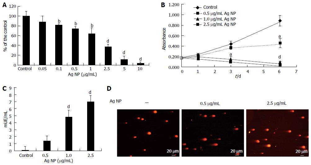
Figure 1 Silver nanoparticles are cytotoxic and genotoxic for human dermal microvascular endothelial cells.
(A) Human dermal microvascular endothelial cells (HMEC) were exposed to various concentrations of silver nanoparticles (Ag NP) for 72 h or (B) were treated with different concentrations of Ag NP for different times. MTT assay was then performed. In (A) data are expressed as the percentage of control and in (B) as absorbance. Data represent the mean ± SD deviation of at least three separate experiments in triplicate. P value was calculated vs untreated cells: aP < 0.05, bP < 0.01, dP < 0.001. C: HMEC were treated with 0.5, 1.0 and 2.5 μg/mL Ag NP for 24 h and the LDH leakage assay was performed. Data are expressed as enzyme unit/mL culture medium and represent the mean ± SD of five separate experiments. P value was calculated vs untreated cells: dP < 0.001; D: Comet assay was performed on HMEC treated with Ag NP for 24 h. After staining with ethidium bromide, the slides were analyzed with a fluorescence microscope.
- Citation: Castiglioni S, Caspani C, Cazzaniga A, Maier JA. Short- and long-term effects of silver nanoparticles on human microvascular endothelial cells. World J Biol Chem 2014; 5(4): 457-464
- URL: https://www.wjgnet.com/1949-8454/full/v5/i4/457.htm
- DOI: https://dx.doi.org/10.4331/wjbc.v5.i4.457


