Copyright
©2014 Baishideng Publishing Group Inc.
World J Biol Chem. Nov 26, 2014; 5(4): 398-408
Published online Nov 26, 2014. doi: 10.4331/wjbc.v5.i4.398
Published online Nov 26, 2014. doi: 10.4331/wjbc.v5.i4.398
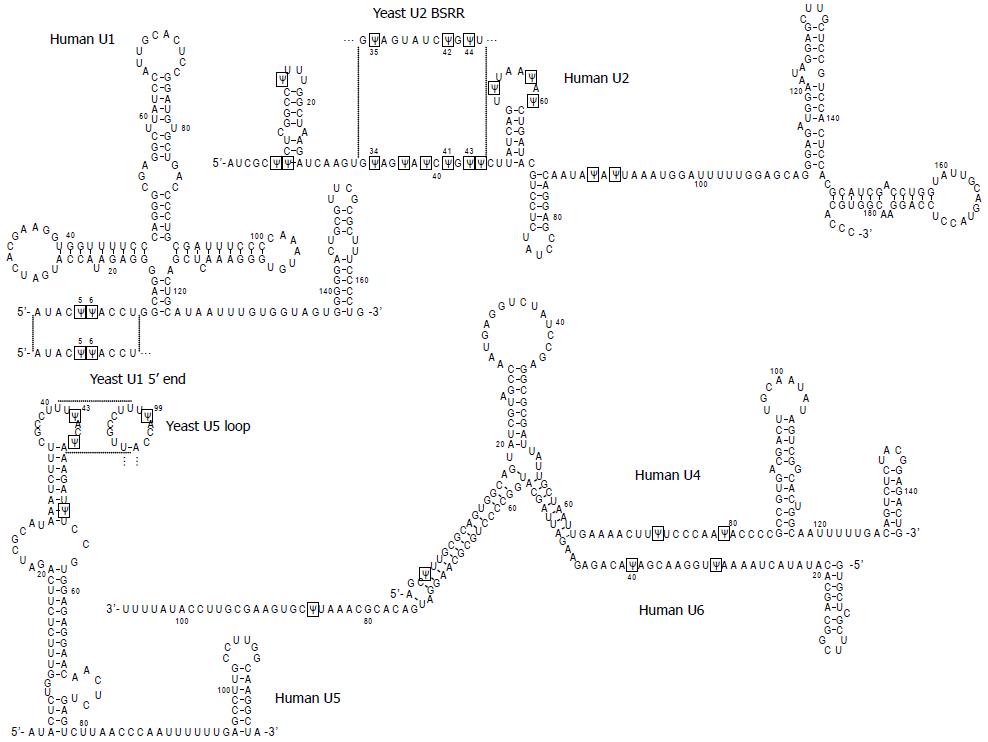
Figure 1 Primary sequences and secondary structures of human spliceosomal snRNAs (U1, U2, U4, U5 and U6).
Pseudouridines (Ψ) are boxed. The sequences of yeast snRNAs where the Ψs have their counterparts in human snRNAs (the 5’ end region of U1, branch site recognition region (BSRR) of U2, and loop region of U5) are also shown. The structures are predicted by the “multifold” program and are consistent with the genetic/biochemical mapping data.
- Citation: Adachi H, Yu YT. Insight into the mechanisms and functions of spliceosomal snRNA pseudouridylation. World J Biol Chem 2014; 5(4): 398-408
- URL: https://www.wjgnet.com/1949-8454/full/v5/i4/398.htm
- DOI: https://dx.doi.org/10.4331/wjbc.v5.i4.398


