Copyright
©2014 Baishideng Publishing Group Inc.
World J Biol Chem. Aug 26, 2014; 5(3): 377-386
Published online Aug 26, 2014. doi: 10.4331/wjbc.v5.i3.377
Published online Aug 26, 2014. doi: 10.4331/wjbc.v5.i3.377
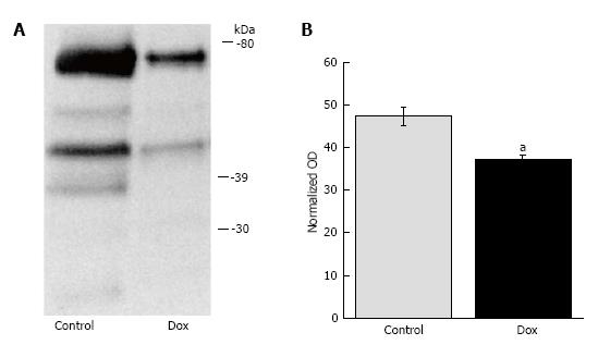
Figure 1 Proteome lysine acetylation status.
A: Representative Western blot showing doxorubicin (Dox)-induced proteome lysine deacetylation; B: Graphical representation of OD units determined by whole lane analysis and normalized to Ponceau staining. aP < 0.050, control vs Dox.
- Citation: Dirks-Naylor AJ, Kouzi SA, Bero JD, Tran NT, Yang S, Mabolo R. Effects of acute doxorubicin treatment on hepatic proteome lysine acetylation status and the apoptotic environment. World J Biol Chem 2014; 5(3): 377-386
- URL: https://www.wjgnet.com/1949-8454/full/v5/i3/377.htm
- DOI: https://dx.doi.org/10.4331/wjbc.v5.i3.377


