Copyright
©2012 Baishideng Publishing Group Co.
World J Biol Chem. May 26, 2012; 3(5): 98-109
Published online May 26, 2012. doi: 10.4331/wjbc.v3.i5.98
Published online May 26, 2012. doi: 10.4331/wjbc.v3.i5.98
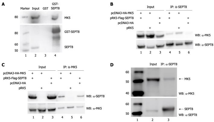
Figure 1 Mitogen-activated protein kinase-activated protein kinase 5 and SEPT8 interact in vitro and in vivo.
A: Glutathione S-transferase (GST) pull-down assay. GST-SEPT8 or GST alone purified from Escherichia coli and immobilized on glutathione-Sepharose beads were incubated for 60 min with lysate of HEK293 cells transfected with GFP-mitogen-activated protein kinase-activated protein kinase 5 (MK5). After washing the beads five times, bound proteins were eluted by boiling, subjected to SDS-PAGE, and immunoblotted with anti-PRAK (upper panel) or anti-SEPT8 (lower panel) antibody. Lane 1: Protein molecular mass marker (in kDa); lane 2: Cell lysate; lane 3: Cell lysate after pull down with GST; lane 4: Cell lysate after pull down with GST-SEPT8; B: Coimmunoprecipitation of FLAG-SEPT8 and hemagglutinin (HA)-MK5. HA-MK5- and/or FLAG-SEPT8-encoding plasmids were transiently expressed in HEK293 cells. Total cellular lysates (input; lanes 1-3) and SEPT8 immunoprecipitates (IP: α-SEPT8; lanes 4-6) were probed with anti-PRAK (upper panel) and anti-SEPT8 (lower panel) antibody. Lane 1: Cell lysate of cells cotransfected with expression plasmids for HA-tagged MK5 and FLAG-tagged SEPT8; lane 2: Cell lysate of cells cotransfected with expression plasmids for HA-tagged MK5 and empty vector for SEPT8; lane 3: Cell lysate of cells cotransfected with expression plasmids for FLAG-tagged SEPT8 and empty vector for MK5; lane 4: Immunoprecipitated lysate of cells cotransfected with expression plasmids for HA-tagged MK5 and FLAG-tagged SEPT8; lane 5: Immunoprecipitated lysate of cells cotransfected with expression plasmids for HA-tagged MK5 and empty vector for SEPT8; lane 6: Immunoprecipitated lysate of cells cotransfected with expression plasmids for FLAG-tagged SEPT8 and empty vector for MK5; C: Coimmunoprecipitation of HA-MK5 and FLAG-SEPT8. HEK293 cells were transiently transfected with expression plasmids for HA-MK5 and/or FLAG-SEPT8. Total cellular lysates (input) and HA-MK5 immunoprecipitates (IP: α-MK5) were probed with anti-SEPT8 (upper panel) and anti-PRAK (lower panel) antibody. Lanes 1-3: Lysates from transfected cells; lanes 4-6: Immunoprecipitation of cell lysates. See (B) for details; D: Coimmunoprecipitation of endogenous MK5 and SEPT8. Endogenous SEPT8 was immunoprecipitated from PC12 cells with anti-SEPT8 antibodies and the precipitate was analyzed for the presence of MK5 by anti-PRAK antibodies. Lane 1: Protein molecular mass marker (in kDa); lane 2: Lysate of PC12 cells (= input); lane 3: Immunoprecipitation with SEPT8 antibodies. The bottom panel shows control western blot with SEPT8 antibodies. IP: Immunoprecipitation; WB: Western blotting.
-
Citation: Shiryaev A, Kostenko S, Dumitriu G, Moens U. Septin 8 is an interaction partner and
in vitro substrate of MK5. World J Biol Chem 2012; 3(5): 98-109 - URL: https://www.wjgnet.com/1949-8454/full/v3/i5/98.htm
- DOI: https://dx.doi.org/10.4331/wjbc.v3.i5.98


