Copyright
©The Author(s) 2023.
World J Biol Chem. Oct 17, 2023; 14(5): 84-98
Published online Oct 17, 2023. doi: 10.4331/wjbc.v14.i5.84
Published online Oct 17, 2023. doi: 10.4331/wjbc.v14.i5.84
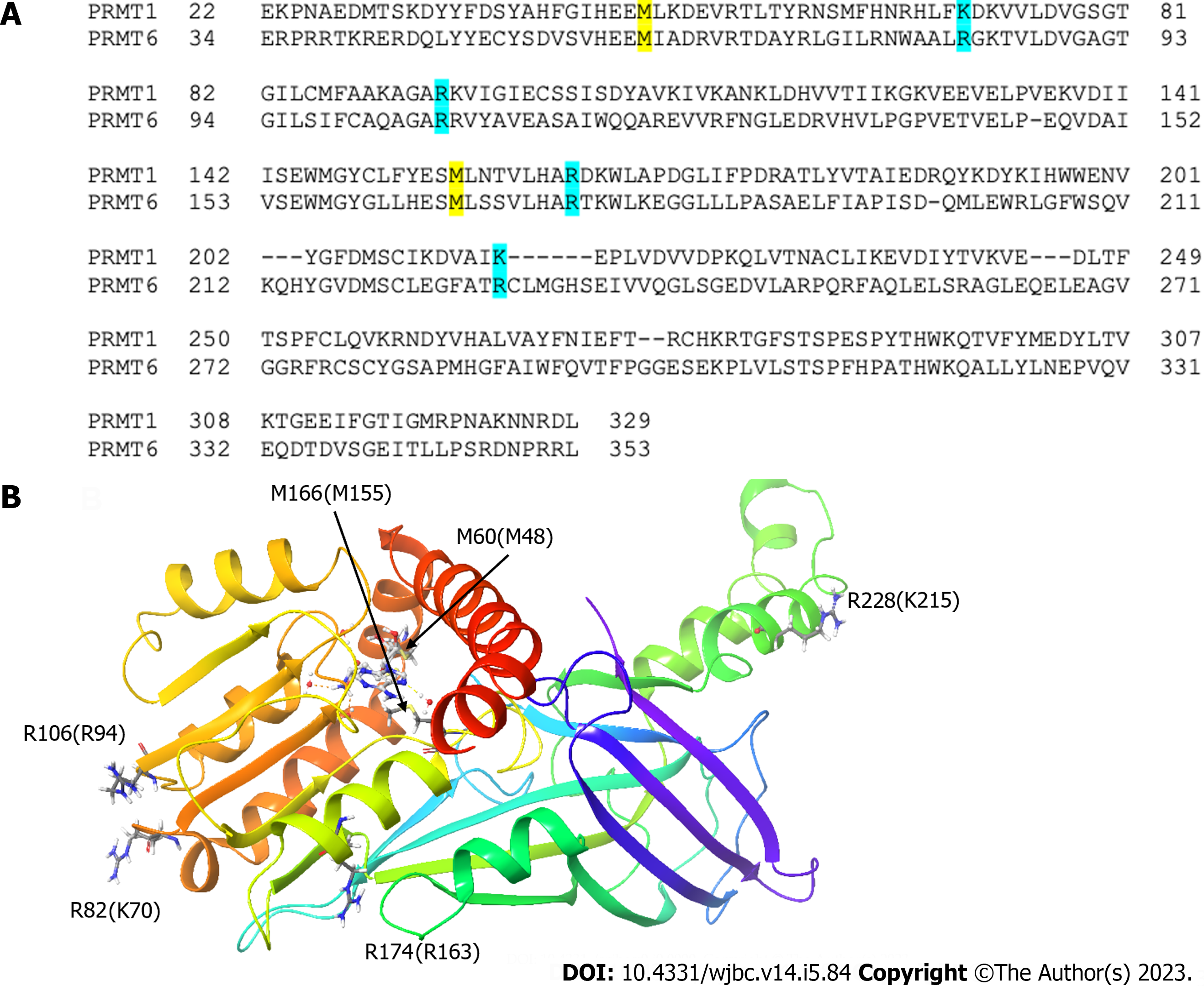
Figure 7 The mutation sites of protein arginine methyltransferase 6 and corresponding residues on protein arginine methyltransferase 1.
A: Sequence alignment of protein arginine methyltransferase (PRMT) 1 and PRMT6 showing the corresponding mutation sites; B: The corresponding mutation sites are highlighted on PRMT6 structure. hPRMT6 monomer (PDB: 4Y30) is shown in a cartoon model. The mutation sites of PRMT6 with their corresponding residues on PRMT1 and the S-Adenosyl-L-homocysteine molecule are shown in a stick model.
- Citation: Cao MT, Feng Y, Zheng YG. Protein arginine methyltransferase 6 is a novel substrate of protein arginine methyltransferase 1. World J Biol Chem 2023; 14(5): 84-98
- URL: https://www.wjgnet.com/1949-8454/full/v14/i5/84.htm
- DOI: https://dx.doi.org/10.4331/wjbc.v14.i5.84


