Copyright
©The Author(s) 2019.
World J Biol Chem. Jan 7, 2019; 10(1): 17-27
Published online Jan 7, 2019. doi: 10.4331/wjbc.v10.i1.17
Published online Jan 7, 2019. doi: 10.4331/wjbc.v10.i1.17
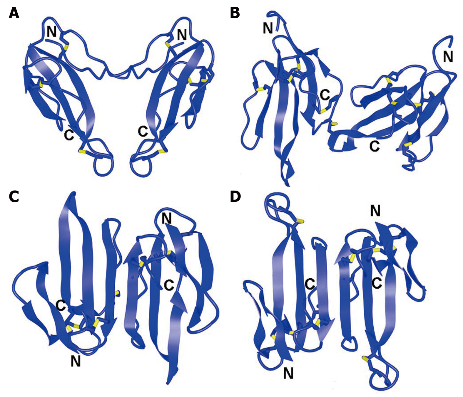
Figure 1 Spatial structures of dimeric three-finger toxins.
A: Homodimer of α-cobratoxin, Protein Data Bank Identification code (PDB ID): 4AEA; B: Irditoxin, PDB ID: 2H7Z; C: Haditoxin, PDB ID: 3HH7; D: κ-bungarotoxin, PDB ID: 1KBA. Disulfide bonds are shown in yellow. N and C indicate N- and C-terminus, respectively.
- Citation: Utkin YN. Last decade update for three-finger toxins: Newly emerging structures and biological activities. World J Biol Chem 2019; 10(1): 17-27
- URL: https://www.wjgnet.com/1949-8454/full/v10/i1/17.htm
- DOI: https://dx.doi.org/10.4331/wjbc.v10.i1.17


