Copyright
©2010 Baishideng Publishing Group Co.
World J Biol Chem. May 26, 2010; 1(5): 95-102
Published online May 26, 2010. doi: 10.4331/wjbc.v1.i5.95
Published online May 26, 2010. doi: 10.4331/wjbc.v1.i5.95
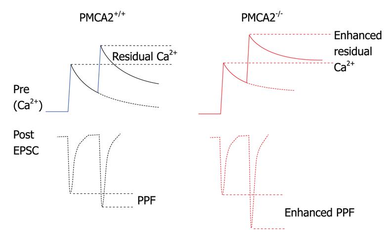
Figure 2 Schematic of the influence of deletion of PMCA2 on “residual” calcium at the PF to PN synapse.
Upper solid traces represent the calcium concentration dynamics in the pre-synaptic terminal in response to a paired stimulation of the PFs; calcium rises once and then again a few milliseconds later. Lower dotted line trace represents the excitatory post synaptic current (EPSC) on the post-synaptic side as a consequence of glutamate released during the rise in calcium in the pre-synaptic terminal; note the larger second EPSC coinciding with the larger peak calcium, the paired pulse facilitation (PPF). In red, in the PMCA2 knockout tissue, calcium rises in the pre-synaptic terminal but then decays more slowly, meaning that on the second stimulation, calcium rises even higher; this is seen as an increase in the peak calcium and also the residual calcium. As a consequence, the second EPSC, dotted lines, lower trace, is larger in amplitude, since more glutamate was released from the PF terminal as a consequence of increased calcium. The recovery of the facilitated EPSCs is also slowed in the PMCA2-/- cerebellum, as residual calcium stays elevated for a longer time in the absence of the PMCA2.
- Citation: Huang H, Nagaraja RY, Garside ML, Akemann W, Knöpfel T, Empson RM. Contribution of plasma membrane Ca2+ ATPase to cerebellar synapse function. World J Biol Chem 2010; 1(5): 95-102
- URL: https://www.wjgnet.com/1949-8454/full/v1/i5/95.htm
- DOI: https://dx.doi.org/10.4331/wjbc.v1.i5.95


