Copyright
©2010 Baishideng Publishing Group Co.
World J Biol Chem. May 26, 2010; 1(5): 109-132
Published online May 26, 2010. doi: 10.4331/wjbc.v1.i5.109
Published online May 26, 2010. doi: 10.4331/wjbc.v1.i5.109
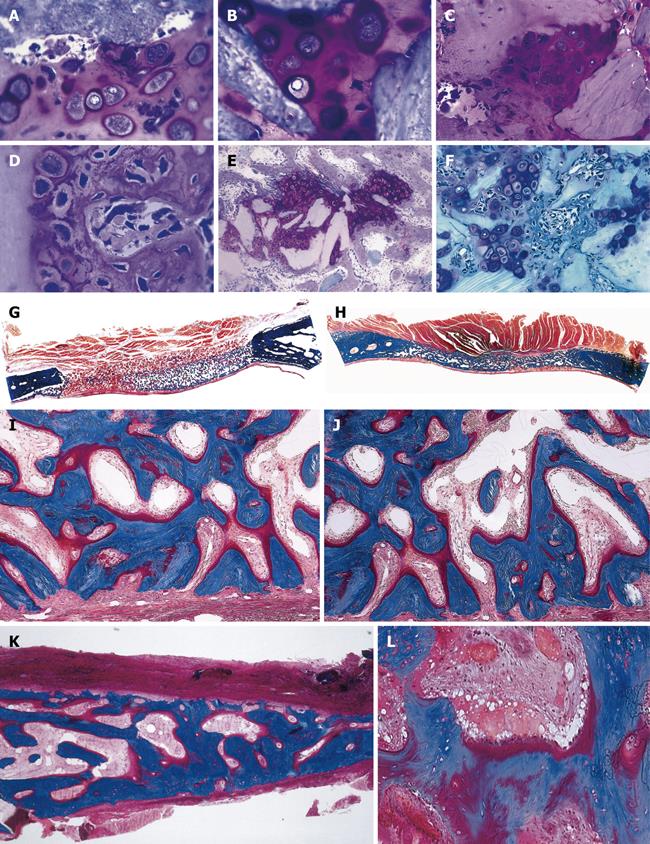
Figure 2 Tissue induction and morphogenesis upon recombination, or reconstitution, of extracted naturally-derived highly purified osteogenic soluble molecular signals with the insoluble signal of the inactive collagenous bone matrix in rodents and non-human primates of the species Papio ursinus (P.
ursinus). A-F: Differentiation of endochondral bone upon implantation of 0.1-0.5 μg osteogenin purified to apparent homogeneity reconstituted with rat insoluble and inactive collagenous bone matrix implanted subcutaneously in rats. Vascular invasion (D, F) initiates chondrolysis and osteoblastic-like cell differentiation by induction attached to the implanted collagenous matrix (F); G-J: Induction of bone formation on days 30 (G, I) and 90 (H, J) by naturally-derived highly purified osteogenic proteins extracted and purified from baboon bone matrices and implanted in non-healing calvarial defects of non-human primates P. ursinus. Mineralized bone (in blue) is surfaced by osteoid seams (red-orange) populated by contiguous osteoblasts; K, L: Highly purified bovine osteogenic proteins additionally purified by heparin-affinity chromatography column induce mineralized bone surfaced by osteoid seams 90 d after implantation in a massive mandibular defect of a human patient (K); high power view (L) shows the mineralized newly formed bone (in blue) surrounding the implanted collagenous matrix as carrier for the osteogenic proteins; this demonstrates the induction of bone formation in the human patient. A-F: Undecalcified section cut at 3 μm stained with toluidine blue after embedding in historesin; G-L: Undecalcified sections cut at 6 μm stained free-floating with a modified Goldner’s trichrome.
- Citation: Ripamonti U. Soluble and insoluble signals sculpt osteogenesis in angiogenesis. World J Biol Chem 2010; 1(5): 109-132
- URL: https://www.wjgnet.com/1949-8454/full/v1/i5/109.htm
- DOI: https://dx.doi.org/10.4331/wjbc.v1.i5.109


