Copyright
©2010 Baishideng Publishing Group Co.
World J Biol Chem. May 26, 2010; 1(5): 109-132
Published online May 26, 2010. doi: 10.4331/wjbc.v1.i5.109
Published online May 26, 2010. doi: 10.4331/wjbc.v1.i5.109
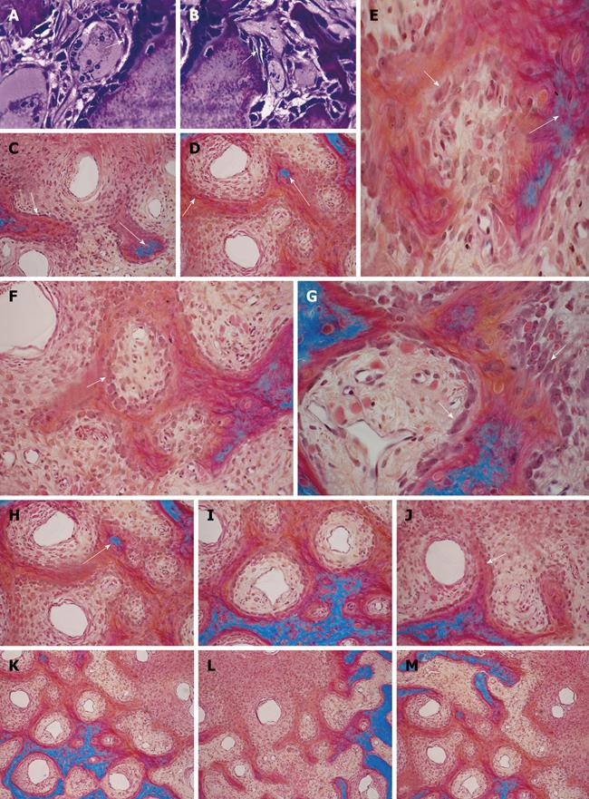
Figure 1 Three-dimensional angiogenesis, capillary sprouting, cellular trafficking and vascular and perivascular stem cell differentiation setting the induction of bone formation and the architecture of the cortico-cancellous osteonic bone.
A, B: Angiogenesis with exquisite intimate relationships between the invading capillaries and the collagenous matrix combined with 0.1-0.5 μg of osteogenin purified to apparent homogeneity from baboon bone matrices after subcutaneous implantation in rodents. Short arrows indicate vascular/perivascular cellular elements migrating from the vascular compartment to the bone forming compartment. The close proximity of the capillary to the newly differentiated osteoblastic cells seemingly provide a continuous flow of osteoprogenitor stem cells differentiating into bone forming cells as soon as pericytic cells move into the bone forming/osteoblastic compartment; C-M: Morphological stages of vascular tissue patterning and morphogenesis of the primate cortico-cancellous bone enveloping the central morphogenetic and osteogenetic vessels (short arrows in C and J). Long arrows point to foci of mineralization within the newly formed mesenchymal condensations with osteoblastic-like cells lining the morphogenetic condensation facing the central blood vessels (short arrow in F). Short arrows in D, E and G indicate osteoblastic-like cells. Mineralized mesenchymal highly cellular condensations (K, L, M) forming and remodeling around central morphogenetic and osteogenetic vessels pattern the induction of osteonic bone formation. Note the intimate relationships of the central blood vessels with osteoblastic/pre-osteoblastic-like cells and other stem cells surfacing the mesenchymal condensations patterning the newly formed osteonic cortico-cancellous bone. Undecalcified sections cut at 6 μm stained free-floating with a modified Goldner’s trichrome.
- Citation: Ripamonti U. Soluble and insoluble signals sculpt osteogenesis in angiogenesis. World J Biol Chem 2010; 1(5): 109-132
- URL: https://www.wjgnet.com/1949-8454/full/v1/i5/109.htm
- DOI: https://dx.doi.org/10.4331/wjbc.v1.i5.109


