Copyright
©The Author(s) 2017.
World J Gastrointest Surg. Feb 27, 2017; 9(2): 68-72
Published online Feb 27, 2017. doi: 10.4240/wjgs.v9.i2.68
Published online Feb 27, 2017. doi: 10.4240/wjgs.v9.i2.68
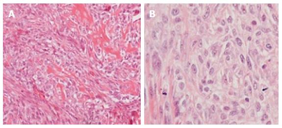
Figure 3 The tumor.
A: The tumor consisted of atypical spindle or polyhedral cells that were intimately associated to neoplastic bone deposited in a lacy pattern (haematoxylin and eosin stain, original magnification × 20); B: The tumor cells were mitotically active and frequently demonstrated atypical mitotic figures (haematoxylin and eosin stain, original magnification × 40).
- Citation: Diamantis A, Christodoulidis G, Vasdeki D, Karasavvidou F, Margonis E, Tepetes K. Giant abdominal osteosarcoma causing intestinal obstruction treated with resection and adjuvant chemotherapy. World J Gastrointest Surg 2017; 9(2): 68-72
- URL: https://www.wjgnet.com/1948-9366/full/v9/i2/68.htm
- DOI: https://dx.doi.org/10.4240/wjgs.v9.i2.68


